Rapid Assessment of Bio-distribution and Antitumor Activity of the Photosensitizer Bremachlorin in a Murine PDAC Model: Detection of PDT-induced Tumor Necrosis by IRDye® 800CW Carboxylate, Using Whole-Body Fluorescent Imaging.
Roisin McMorrow, Henriette S de Bruijn, Ivo Que, Debra C Stuurman, Corrina M A de Ridder, Michail Doukas, Dominic J Robinson, Laura Mezzanotte, Clemens W G M Lowik
{"title":"Rapid Assessment of Bio-distribution and Antitumor Activity of the Photosensitizer Bremachlorin in a Murine PDAC Model: Detection of PDT-induced Tumor Necrosis by IRDye® 800CW Carboxylate, Using Whole-Body Fluorescent Imaging.","authors":"Roisin McMorrow, Henriette S de Bruijn, Ivo Que, Debra C Stuurman, Corrina M A de Ridder, Michail Doukas, Dominic J Robinson, Laura Mezzanotte, Clemens W G M Lowik","doi":"10.1007/s11307-024-01921-1","DOIUrl":null,"url":null,"abstract":"<p><p>Photodynamic therapy (PDT) is a light-based anticancer therapy that can induce tumor necrosis and/or apoptosis. Two important factors contributing to the efficacy of PDT are the concentration of the photosensitizer in the tumor tissue and its preferential accumulation in the tumor tissue compared to that in normal tissues. In this study, we investigated the use of optical imaging for monitoring whole-body bio-distribution of the fluorescent (660 nm) photosensitizer Bremachlorin in vivo, in a murine pancreatic ductal adenocarcinoma (PDAC) model. Moreover, we non-invasively, examined the induction of tumor necrosis after PDT treatment using near-infrared fluorescent imaging of the necrosis avid cyanine dye IRDye®-800CW Carboxylate. Using whole-body fluorescence imaging, we observed that Bremachlorin preferentially accumulated in pancreatic tumors. Furthermore, in a longitudinal study we showed that 3 hours after Bremachlorin administration, the fluorescent tumor signal reached its maximum. In addition, the tumor-to-background ratio at all-time points was approximately 1.4. Ex vivo, at 6 hours after Bremachlorin administration, the tumor-to-muscle or -normal pancreas ratio exhibited a greater difference than it did at 24 hours, suggesting that, in terms of efficacy, 6 hours after Bremachlorin administration was an effective time point for PDT treatment of PDAC. In vivo administration of the near infrared fluorescence agent IRDye®-800CW Carboxylate showed that PDT, 6 hours after administration of Bremachlorin, selectively induced necrosis in the tumor tissues, which was subsequently confirmed histologically. In conclusion, by using in vivo fluorescence imaging, we could non-invasively and longitudinally monitor, the whole-body distribution of Bremachlorin. Furthermore, we successfully used IRDye®-800CW Carboxylate, a near-infrared fluorescent necrosis avid agent, to image PDT-induced necrotic cell death as a measure of therapeutic efficacy. This study showed how fluorescence can be applied for optimizing, and assessing the efficacy of, PDT.</p>","PeriodicalId":18760,"journal":{"name":"Molecular Imaging and Biology","volume":" ","pages":"616-627"},"PeriodicalIF":2.5000,"publicationDate":"2024-08-01","publicationTypes":"Journal Article","fieldsOfStudy":null,"isOpenAccess":false,"openAccessPdf":"https://www.ncbi.nlm.nih.gov/pmc/articles/PMC11281978/pdf/","citationCount":"0","resultStr":null,"platform":"Semanticscholar","paperid":null,"PeriodicalName":"Molecular Imaging and Biology","FirstCategoryId":"3","ListUrlMain":"https://doi.org/10.1007/s11307-024-01921-1","RegionNum":4,"RegionCategory":"医学","ArticlePicture":[],"TitleCN":null,"AbstractTextCN":null,"PMCID":null,"EPubDate":"2024/6/18 0:00:00","PubModel":"Epub","JCR":"Q2","JCRName":"RADIOLOGY, NUCLEAR MEDICINE & MEDICAL IMAGING","Score":null,"Total":0}
引用次数: 0
Abstract
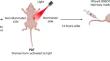
Photodynamic therapy (PDT) is a light-based anticancer therapy that can induce tumor necrosis and/or apoptosis. Two important factors contributing to the efficacy of PDT are the concentration of the photosensitizer in the tumor tissue and its preferential accumulation in the tumor tissue compared to that in normal tissues. In this study, we investigated the use of optical imaging for monitoring whole-body bio-distribution of the fluorescent (660 nm) photosensitizer Bremachlorin in vivo, in a murine pancreatic ductal adenocarcinoma (PDAC) model. Moreover, we non-invasively, examined the induction of tumor necrosis after PDT treatment using near-infrared fluorescent imaging of the necrosis avid cyanine dye IRDye®-800CW Carboxylate. Using whole-body fluorescence imaging, we observed that Bremachlorin preferentially accumulated in pancreatic tumors. Furthermore, in a longitudinal study we showed that 3 hours after Bremachlorin administration, the fluorescent tumor signal reached its maximum. In addition, the tumor-to-background ratio at all-time points was approximately 1.4. Ex vivo, at 6 hours after Bremachlorin administration, the tumor-to-muscle or -normal pancreas ratio exhibited a greater difference than it did at 24 hours, suggesting that, in terms of efficacy, 6 hours after Bremachlorin administration was an effective time point for PDT treatment of PDAC. In vivo administration of the near infrared fluorescence agent IRDye®-800CW Carboxylate showed that PDT, 6 hours after administration of Bremachlorin, selectively induced necrosis in the tumor tissues, which was subsequently confirmed histologically. In conclusion, by using in vivo fluorescence imaging, we could non-invasively and longitudinally monitor, the whole-body distribution of Bremachlorin. Furthermore, we successfully used IRDye®-800CW Carboxylate, a near-infrared fluorescent necrosis avid agent, to image PDT-induced necrotic cell death as a measure of therapeutic efficacy. This study showed how fluorescence can be applied for optimizing, and assessing the efficacy of, PDT.
期刊介绍:
Molecular Imaging and Biology (MIB) invites original contributions (research articles, review articles, commentaries, etc.) on the utilization of molecular imaging (i.e., nuclear imaging, optical imaging, autoradiography and pathology, MRI, MPI, ultrasound imaging, radiomics/genomics etc.) to investigate questions related to biology and health. The objective of MIB is to provide a forum to the discovery of molecular mechanisms of disease through the use of imaging techniques. We aim to investigate the biological nature of disease in patients and establish new molecular imaging diagnostic and therapy procedures.
Some areas that are covered are:
Preclinical and clinical imaging of macromolecular targets (e.g., genes, receptors, enzymes) involved in significant biological processes.
The design, characterization, and study of new molecular imaging probes and contrast agents for the functional interrogation of macromolecular targets.
Development and evaluation of imaging systems including instrumentation, image reconstruction algorithms, image analysis, and display.
Development of molecular assay approaches leading to quantification of the biological information obtained in molecular imaging.
Study of in vivo animal models of disease for the development of new molecular diagnostics and therapeutics.
Extension of in vitro and in vivo discoveries using disease models, into well designed clinical research investigations.
Clinical molecular imaging involving clinical investigations, clinical trials and medical management or cost-effectiveness studies.


 分享
分享

 求助内容:
求助内容: 应助结果提醒方式:
 扫码关注我们
