Sydney L Olson, Razeen J Akbar, Adrianna Gorniak, Laura I Fuhr, Mostafa A Borahay
{"title":"Hypoxia in uterine fibroids: role in pathobiology and therapeutic opportunities.","authors":"Sydney L Olson, Razeen J Akbar, Adrianna Gorniak, Laura I Fuhr, Mostafa A Borahay","doi":"10.3390/oxygen4020013","DOIUrl":null,"url":null,"abstract":"<p><p>Uterine fibroids are the most common tumors in females affecting up to 70% of women world-wide, yet targeted therapeutic options are limited. Oxidative stress has recently surfaced as a key driver of fibroid pathogenesis and provides insights into hypoxia-induced cell transformation, extracellular matrix pathophysiology, hypoxic cell signaling cascades, and uterine biology. Hypoxia drives fibroid tumorigenesis through (1) promoting myometrial stem cell proliferation, (2) causing DNA damage propelling transformation of stem cells to tumor initiating cells, and (3) driving excess extracellular matrix (ECM) production. Common fibroid-associated DNA mutations include MED12 mutations, HMGA2 overexpression, and Fumarate hydratase loss of function. Evidence suggests an interaction between hypoxia signaling and these mutations. Fibroid development and growth are promoted by hypoxia-triggered cell signaling via various pathways including HIF-1, TGFβ, and Wnt/β-catenin. Fibroid-associated hypoxia persists due to antioxidant imbalance, ECM accumulation, and growth beyond adequate vascular supply. Current clinically available fibroid treatments do not take advantage of hypoxia-targeting therapies. Growing pre-clinical and clinical studies identify ROS inhibitors, anti-HIF-1 agents, Wnt/β-catenin inhibition, and TGFβ cascade inhibitors as agents that may reduce fibroid development and growth through targeting hypoxia.</p>","PeriodicalId":74387,"journal":{"name":"Oxygen (Basel, Switzerland)","volume":"4 2","pages":"236-252"},"PeriodicalIF":0.0000,"publicationDate":"2024-06-01","publicationTypes":"Journal Article","fieldsOfStudy":null,"isOpenAccess":false,"openAccessPdf":"https://www.ncbi.nlm.nih.gov/pmc/articles/PMC11218552/pdf/","citationCount":"0","resultStr":null,"platform":"Semanticscholar","paperid":null,"PeriodicalName":"Oxygen (Basel, Switzerland)","FirstCategoryId":"1085","ListUrlMain":"https://doi.org/10.3390/oxygen4020013","RegionNum":0,"RegionCategory":null,"ArticlePicture":[],"TitleCN":null,"AbstractTextCN":null,"PMCID":null,"EPubDate":"2024/5/28 0:00:00","PubModel":"Epub","JCR":"","JCRName":"","Score":null,"Total":0}
引用次数: 0
Abstract
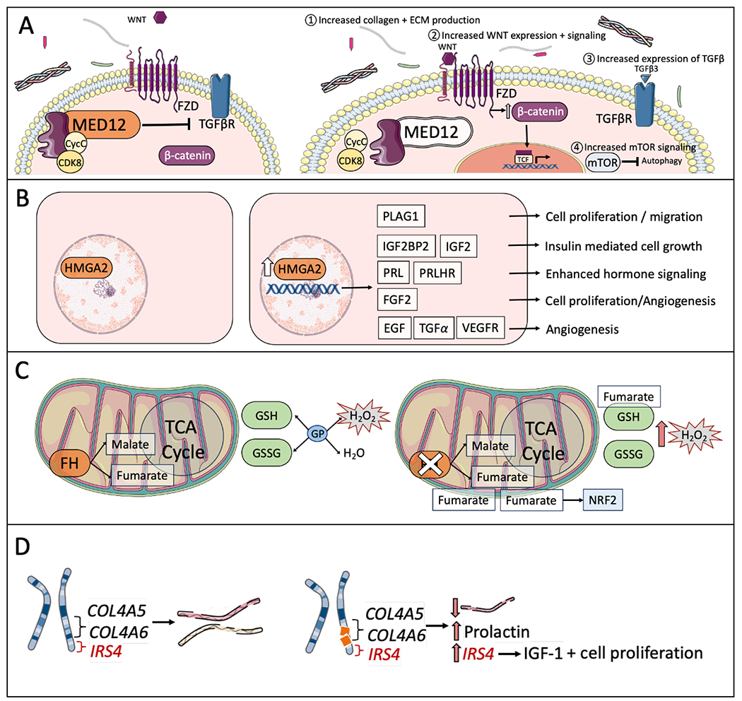
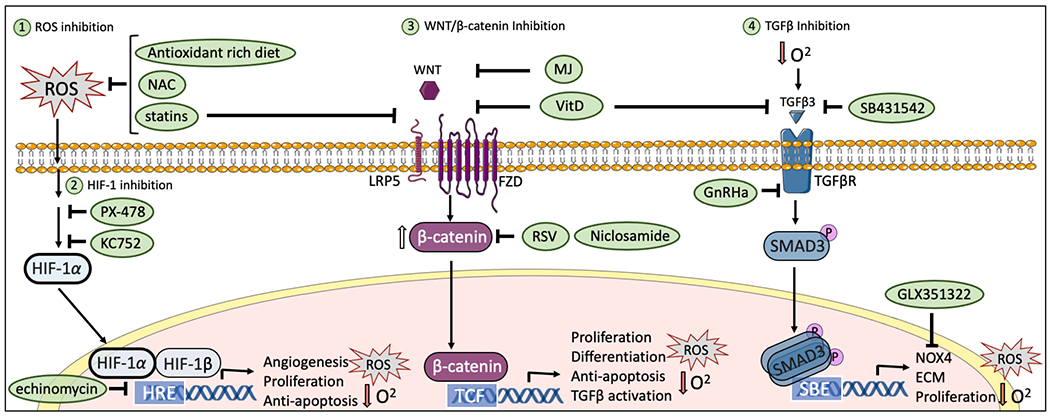
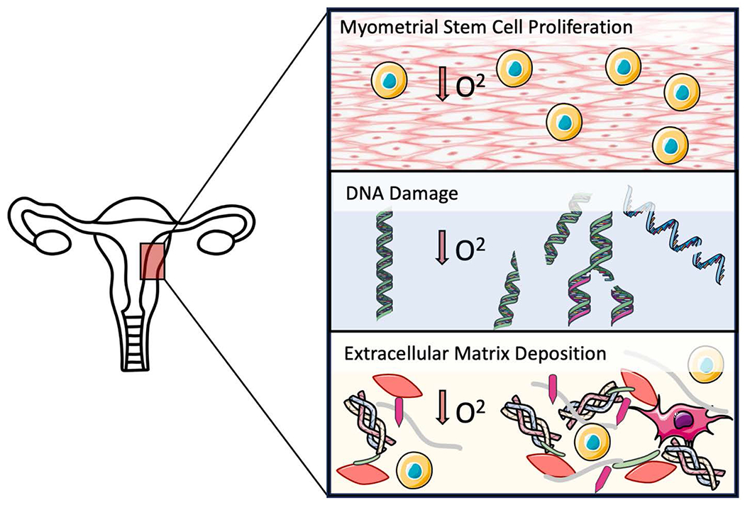
Uterine fibroids are the most common tumors in females affecting up to 70% of women world-wide, yet targeted therapeutic options are limited. Oxidative stress has recently surfaced as a key driver of fibroid pathogenesis and provides insights into hypoxia-induced cell transformation, extracellular matrix pathophysiology, hypoxic cell signaling cascades, and uterine biology. Hypoxia drives fibroid tumorigenesis through (1) promoting myometrial stem cell proliferation, (2) causing DNA damage propelling transformation of stem cells to tumor initiating cells, and (3) driving excess extracellular matrix (ECM) production. Common fibroid-associated DNA mutations include MED12 mutations, HMGA2 overexpression, and Fumarate hydratase loss of function. Evidence suggests an interaction between hypoxia signaling and these mutations. Fibroid development and growth are promoted by hypoxia-triggered cell signaling via various pathways including HIF-1, TGFβ, and Wnt/β-catenin. Fibroid-associated hypoxia persists due to antioxidant imbalance, ECM accumulation, and growth beyond adequate vascular supply. Current clinically available fibroid treatments do not take advantage of hypoxia-targeting therapies. Growing pre-clinical and clinical studies identify ROS inhibitors, anti-HIF-1 agents, Wnt/β-catenin inhibition, and TGFβ cascade inhibitors as agents that may reduce fibroid development and growth through targeting hypoxia.


 分享
分享

 求助内容:
求助内容: 应助结果提醒方式:
 扫码关注我们
