{"title":"Extracellular vesicles derived from \"serum and glucose\" deprived HUCMSCs promoted skin wound healing through enhanced angiogenesis.","authors":"Xiaopeng Wu, Pingping Yuan, Na Wei, Chaoqun Ma, Mingdi Fu, Wei Wu","doi":"10.1007/s11010-024-05058-1","DOIUrl":null,"url":null,"abstract":"<p><p>Extracellular vesicles (EVs) produced from MSCs were currently considered as a novel therapeutic agent for skin tissue regeneration and repair. Preconditioning stem cells may activate more molecular pathways and release more bioactive agents. In this study, we obtained EVs from normal (N-EVs) and serum- and glucose-deprived (SGD-EVs) human umbilical cord mesenchymal stem cells (HUCMSCs), and showed that SGD-EVs promoted the migration, proliferation, and tube formation of HUVECs in vitro. In vivo experiments utilizing a rat model show that both N-EVs and SGD-EVs boosted angiogenesis of skin defects and accelerated skin wound healing, while treating wounds with SGD-EVs led to faster skin healing and enhanced angiogenesis. miRNA sequencing showed that miR-29a-3p was abundant in SGD-EVs, and overexpressing miR-29a-3p enhanced the angiogenic ability of HUVECs, while inhibiting miR-29a-3p presented the opposite effect. Further studies demonstrated that miR-29a-3p directly targeted CTNNBIP1, which mediated angiogenesis of HUCMSCs-derived EVs through inhibiting CTNNBIP1 to activate Wnt/β-catenin signaling pathway. Taken together, these findings suggested that SGD-EVs promote angiogenesis via transferring miR-29a-3p, and activation of Wnt/β-catenin signaling pathway played a crucial role in SGD-EVs-induced VEGFA production during wound angiogenesis. Our results offered a new avenue for modifying EVs to enhance tissue angiogenesis and augment its role in skin repair.</p>","PeriodicalId":18724,"journal":{"name":"Molecular and Cellular Biochemistry","volume":" ","pages":"1255-1273"},"PeriodicalIF":3.7000,"publicationDate":"2025-02-01","publicationTypes":"Journal Article","fieldsOfStudy":null,"isOpenAccess":false,"openAccessPdf":"","citationCount":"0","resultStr":null,"platform":"Semanticscholar","paperid":null,"PeriodicalName":"Molecular and Cellular Biochemistry","FirstCategoryId":"99","ListUrlMain":"https://doi.org/10.1007/s11010-024-05058-1","RegionNum":2,"RegionCategory":"生物学","ArticlePicture":[],"TitleCN":null,"AbstractTextCN":null,"PMCID":null,"EPubDate":"2024/7/5 0:00:00","PubModel":"Epub","JCR":"Q3","JCRName":"CELL BIOLOGY","Score":null,"Total":0}
引用次数: 0
Abstract
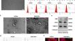
Extracellular vesicles (EVs) produced from MSCs were currently considered as a novel therapeutic agent for skin tissue regeneration and repair. Preconditioning stem cells may activate more molecular pathways and release more bioactive agents. In this study, we obtained EVs from normal (N-EVs) and serum- and glucose-deprived (SGD-EVs) human umbilical cord mesenchymal stem cells (HUCMSCs), and showed that SGD-EVs promoted the migration, proliferation, and tube formation of HUVECs in vitro. In vivo experiments utilizing a rat model show that both N-EVs and SGD-EVs boosted angiogenesis of skin defects and accelerated skin wound healing, while treating wounds with SGD-EVs led to faster skin healing and enhanced angiogenesis. miRNA sequencing showed that miR-29a-3p was abundant in SGD-EVs, and overexpressing miR-29a-3p enhanced the angiogenic ability of HUVECs, while inhibiting miR-29a-3p presented the opposite effect. Further studies demonstrated that miR-29a-3p directly targeted CTNNBIP1, which mediated angiogenesis of HUCMSCs-derived EVs through inhibiting CTNNBIP1 to activate Wnt/β-catenin signaling pathway. Taken together, these findings suggested that SGD-EVs promote angiogenesis via transferring miR-29a-3p, and activation of Wnt/β-catenin signaling pathway played a crucial role in SGD-EVs-induced VEGFA production during wound angiogenesis. Our results offered a new avenue for modifying EVs to enhance tissue angiogenesis and augment its role in skin repair.
期刊介绍:
Molecular and Cellular Biochemistry: An International Journal for Chemical Biology in Health and Disease publishes original research papers and short communications in all areas of the biochemical sciences, emphasizing novel findings relevant to the biochemical basis of cellular function and disease processes, as well as the mechanics of action of hormones and chemical agents. Coverage includes membrane transport, receptor mechanism, immune response, secretory processes, and cytoskeletal function, as well as biochemical structure-function relationships in the cell.
In addition to the reports of original research, the journal publishes state of the art reviews. Specific subjects covered by Molecular and Cellular Biochemistry include cellular metabolism, cellular pathophysiology, enzymology, ion transport, lipid biochemistry, membrane biochemistry, molecular biology, nuclear structure and function, and protein chemistry.


 分享
分享
 求助内容:
求助内容: 应助结果提醒方式:
 扫码关注我们
