Deep learning‐based prediction of the dose–volume histograms for volumetric modulated arc therapy of left‐sided breast cancer

IF 3.2 2区 医学 Q1 RADIOLOGY, NUCLEAR MEDICINE & MEDICAL IMAGING Medical physics Pub Date : 2024-09-18 DOI:10.1002/mp.17410
Akseli Leino, Janne Heikkilä, Tuomas Virén, Juuso T. J. Honkanen, Jan Seppälä, Henri Korkalainen
{"title":"Deep learning‐based prediction of the dose–volume histograms for volumetric modulated arc therapy of left‐sided breast cancer","authors":"Akseli Leino, Janne Heikkilä, Tuomas Virén, Juuso T. J. Honkanen, Jan Seppälä, Henri Korkalainen","doi":"10.1002/mp.17410","DOIUrl":null,"url":null,"abstract":"BackgroundThe advancements in artificial intelligence and computational power have made deep learning an attractive tool for radiotherapy treatment planning. Deep learning has the potential to significantly simplify the trial‐and‐error process involved in inverse planning required by modern treatment techniques such as volumetric modulated arc therapy (VMAT). In this study, we explore the ability of deep learning to predict organ‐at‐risk (OAR) dose–volume histograms (DVHs) of left‐sided breast cancer patients undergoing VMAT treatment based solely on their anatomical characteristics. The predicted DVHs could be used to derive patient‐specific dose constraints and dose objectives, streamlining the treatment planning process, standardizing the quality of the plans, and personalizing the treatment planning.PurposeThis study aimed to develop a deep learning‐based framework for the prediction of organ‐specific dose–volume histograms (DVH) based on structures delineated for left‐sided breast cancer treatment.MethodsWe used a dataset of 249 left‐sided breast cancer patients treated with tangential VMAT fields. We extracted delineated structures and dose distributions for each patient and derived slice‐by‐slice DVHs for planning target volume (PTV) and organs‐at‐risk. The patients were divided into training (70%, <jats:italic>n </jats:italic>= 174), validation (10%, <jats:italic>n </jats:italic>= 24), and test (20%, <jats:italic>n </jats:italic>= 51) sets. Collected data were used to train a deep learning model for the prediction of the DVHs based on the delineated structures. The developed deep learning model comprised a modified DenseNet architecture followed by a recurrent neural network.ResultsIn the independent test set (<jats:italic>n</jats:italic> = 51), the point‐wise differences in the slice‐by‐slice DVHs between the clinical and predicted DVHs were small; the mean squared errors were 3.53, 1.58, 2.28, 3.37, and 1.44 [×10<jats:sup>−4</jats:sup>] for PTV, heart, ipsilateral lung, contralateral lung, and contralateral breast, respectively. With the derived cumulative DVHs, the mean absolute difference ± standard deviation of mean doses between the clinical and the predicted DVH were 0.08 ± 0.04 Gy, 0.24 ± 0.22 Gy, 0.73 ± 0.46 Gy, 0.07 ± 0.06 Gy, and 0.14 ± 0.14 Gy for PTV, heart, ipsilateral lung, contralateral lung, and contralateral breast, respectively.ConclusionsThe deep learning‐based approach enabled automatic and reliable prediction of the DVH based on delineated structures. The predicted DVHs could potentially serve as patient‐specific clinical goals used to aid treatment planning and avoid suboptimal plans or to derive optimization objectives and constraints for automated treatment planning.","PeriodicalId":18384,"journal":{"name":"Medical physics","volume":null,"pages":null},"PeriodicalIF":3.2000,"publicationDate":"2024-09-18","publicationTypes":"Journal Article","fieldsOfStudy":null,"isOpenAccess":false,"openAccessPdf":"","citationCount":"0","resultStr":null,"platform":"Semanticscholar","paperid":null,"PeriodicalName":"Medical physics","FirstCategoryId":"3","ListUrlMain":"https://doi.org/10.1002/mp.17410","RegionNum":2,"RegionCategory":"医学","ArticlePicture":[],"TitleCN":null,"AbstractTextCN":null,"PMCID":null,"EPubDate":"","PubModel":"","JCR":"Q1","JCRName":"RADIOLOGY, NUCLEAR MEDICINE & MEDICAL IMAGING","Score":null,"Total":0}
引用次数: 0

Abstract

BackgroundThe advancements in artificial intelligence and computational power have made deep learning an attractive tool for radiotherapy treatment planning. Deep learning has the potential to significantly simplify the trial‐and‐error process involved in inverse planning required by modern treatment techniques such as volumetric modulated arc therapy (VMAT). In this study, we explore the ability of deep learning to predict organ‐at‐risk (OAR) dose–volume histograms (DVHs) of left‐sided breast cancer patients undergoing VMAT treatment based solely on their anatomical characteristics. The predicted DVHs could be used to derive patient‐specific dose constraints and dose objectives, streamlining the treatment planning process, standardizing the quality of the plans, and personalizing the treatment planning.PurposeThis study aimed to develop a deep learning‐based framework for the prediction of organ‐specific dose–volume histograms (DVH) based on structures delineated for left‐sided breast cancer treatment.MethodsWe used a dataset of 249 left‐sided breast cancer patients treated with tangential VMAT fields. We extracted delineated structures and dose distributions for each patient and derived slice‐by‐slice DVHs for planning target volume (PTV) and organs‐at‐risk. The patients were divided into training (70%, n = 174), validation (10%, n = 24), and test (20%, n = 51) sets. Collected data were used to train a deep learning model for the prediction of the DVHs based on the delineated structures. The developed deep learning model comprised a modified DenseNet architecture followed by a recurrent neural network.ResultsIn the independent test set (n = 51), the point‐wise differences in the slice‐by‐slice DVHs between the clinical and predicted DVHs were small; the mean squared errors were 3.53, 1.58, 2.28, 3.37, and 1.44 [×10−4] for PTV, heart, ipsilateral lung, contralateral lung, and contralateral breast, respectively. With the derived cumulative DVHs, the mean absolute difference ± standard deviation of mean doses between the clinical and the predicted DVH were 0.08 ± 0.04 Gy, 0.24 ± 0.22 Gy, 0.73 ± 0.46 Gy, 0.07 ± 0.06 Gy, and 0.14 ± 0.14 Gy for PTV, heart, ipsilateral lung, contralateral lung, and contralateral breast, respectively.ConclusionsThe deep learning‐based approach enabled automatic and reliable prediction of the DVH based on delineated structures. The predicted DVHs could potentially serve as patient‐specific clinical goals used to aid treatment planning and avoid suboptimal plans or to derive optimization objectives and constraints for automated treatment planning.
查看原文
分享 分享
微信好友 朋友圈 QQ好友 复制链接
本刊更多论文
基于深度学习的左侧乳腺癌容积调制弧治疗剂量-容积直方图预测
背景人工智能和计算能力的进步已使深度学习成为放疗治疗计划中极具吸引力的工具。深度学习有可能大大简化现代治疗技术(如容积调制弧治疗(VMAT))所需的反向规划试错过程。在本研究中,我们探索了深度学习预测接受 VMAT 治疗的左侧乳腺癌患者的风险器官(OAR)剂量-体积直方图(DVH)的能力。预测的剂量容积直方图可用于推导患者特异性剂量约束和剂量目标,从而简化治疗计划流程,实现计划质量标准化和治疗计划个性化。Purpose This study aimed to develop a deep learning-based framework for the prediction of organ-specific dose-volume histograms (DVH) based on structures delineated for left-sided breast cancer treatment.Methods 
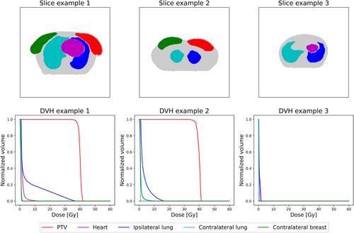
We used a dataset of 249 left-sided breast cancer patients treated with tangential VMAT fields.我们使用了249名接受切向VMAT场治疗的左侧乳腺癌患者的数据集。我们提取了每位患者的划定结构和剂量分布,并得出了规划靶体积(PTV)和危险器官的逐片 DVH。患者被分为训练集(70%,n = 174)、验证集(10%,n = 24)和测试集(20%,n = 51)。收集到的数据被用于训练深度学习模型,以便根据划定的结构预测 DVH。结果在独立测试集(n = 51)中,临床 DVH 与预测 DVH 之间的逐片 DVH 点差异很小;PTV、心脏、同侧肺、对侧肺和对侧乳腺的平均平方误差分别为 3.53、1.58、2.28、3.37 和 1.44 [×10-4]。根据得出的累积DVHs,PTV、心脏、同侧肺、对侧肺和对侧乳房的临床剂量与预测DVH之间的平均剂量绝对差值±标准偏差分别为0.08±0.04 Gy、0.24±0.22 Gy、0.73±0.46 Gy、0.07±0.06 Gy和0.14±0.14 Gy。预测出的 DVH 有可能作为特定患者的临床目标,用于辅助治疗计划和避免次优计划,或用于推导自动治疗计划的优化目标和约束条件。
本文章由计算机程序翻译,如有差异,请以英文原文为准。
求助全文
约1分钟内获得全文 去求助
来源期刊
Medical physics
Medical physics 医学-核医学
CiteScore
6.80
自引率
15.80%
发文量
660
审稿时长
1.7 months
期刊介绍: Medical Physics publishes original, high impact physics, imaging science, and engineering research that advances patient diagnosis and therapy through contributions in 1) Basic science developments with high potential for clinical translation 2) Clinical applications of cutting edge engineering and physics innovations 3) Broadly applicable and innovative clinical physics developments Medical Physics is a journal of global scope and reach. By publishing in Medical Physics your research will reach an international, multidisciplinary audience including practicing medical physicists as well as physics- and engineering based translational scientists. We work closely with authors of promising articles to improve their quality.
期刊最新文献
Individual curved‐needle interstitial template created using three‐dimensional printing for brachytherapy for distal parauterine tumor recurrence Rapid CNN‐based needle localization for automatic slice alignment in MR‐guided interventions using 3D undersampled radial white‐marker imaging Long‐term beam output stability of an accelerator‐based boron neutron capture therapy system Optimization of reconstruction in quantitative brain PET images: Benefits from PSF modeling and correction of edge artifacts Deep learning‐based prediction of the dose–volume histograms for volumetric modulated arc therapy of left‐sided breast cancer
×
引用
GB/T 7714-2015
复制
MLA
复制
APA
复制
导出至
BibTeX EndNote RefMan NoteFirst NoteExpress
×
×
提示
您的信息不完整,为了账户安全,请先补充。
现在去补充
×
提示
您因"违规操作"
具体请查看互助需知
我知道了
×
提示
现在去查看 取消
×
提示
确定
0
微信
客服QQ
Book学术公众号 扫码关注我们
反馈
×
意见反馈
请填写您的意见或建议
请填写您的手机或邮箱
已复制链接
已复制链接
快去分享给好友吧!
我知道了
×
扫码分享
扫码分享
Book学术官方微信
Book学术文献互助
Book学术文献互助群
群 号:481959085
Book学术
文献互助 智能选刊 最新文献 互助须知 联系我们:info@booksci.cn
Book学术提供免费学术资源搜索服务,方便国内外学者检索中英文文献。致力于提供最便捷和优质的服务体验。
Copyright © 2023 Book学术 All rights reserved.
ghs 京公网安备 11010802042870号 京ICP备2023020795号-1