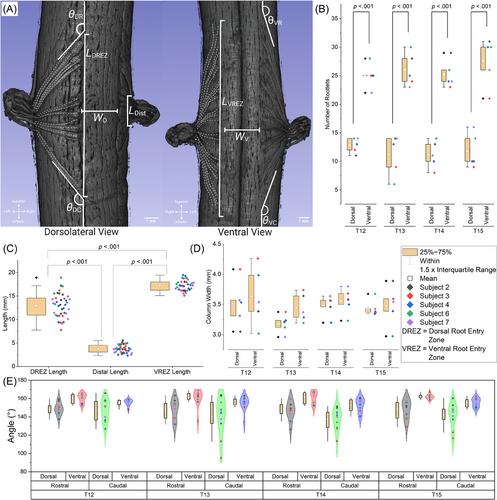
Spinal cord stimulation (SCS) is approved by the Food and Drug Administration for treating chronic intractable pain in the back, trunk, or limbs through stimulation of the dorsal column. Numerous studies have used swine as an analog of the human spinal cord to better understand SCS and further improve its efficacy. We performed high-resolution imaging of the porcine spinal cord with intact dura mater using micro-computed tomography (μCT) to construct detailed 3-dimensional (3D) visualizations of the spinal cord and characterize the morphology of the dorsal and ventral rootlets.
We obtained spinal cords from Yorkshire/Landrace crossbred swine (N = 7), stained samples with osmium tetroxide, and performed μCT imaging of the T12-T15 levels at isotropic voxel resolutions ranging from 3.3 to 50 μm. We measured the anatomical morphology using the 3D volumes and compared our results to measurements previously collected from swine and human spinal cords via microdissection techniques in prior literature.
While the porcine thoracic-lumbar spinal cord is a popular model for SCS, we highlight multiple notable differences compared to previously published T8-T12 human measurements including rootlet counts (porcine dorsal/ventral: 12.2 ± 2.6, 26.6 ± 3.4; human dorsal/ventral: 5.3 ± 1.3, 4.4 ± 2.4), rootlet angles (porcine ventral-rostral: 161 ± 1°, ventral-caudal: 155 ± 6°, dorsal-rostral: 148 ± 9°, dorsal-caudal: 142 ± 6°; human ventral-rostral: 170 ± 3°, ventral-caudal: 22 ± 10°, dorsal-rostral: 171 ± 3°, dorsal-caudal: 15 ± 7°), and the presence and count of dorsal rootlet bundles.
Detailed measurements and highlighted differences between human and porcine spinal cords can inform variations in modeling and electrophysiological experiments between the two species. In contrast to other approaches for measuring the spinal cord and rootlet morphology, our method keeps the dura intact, reducing potential artifacts from dissection.


 分享
分享
 求助内容:
求助内容: 应助结果提醒方式:
 扫码关注我们
