Tanja Kuhm, Clémence Taisne, Cecilia de Agrela Pinto, Luca Gross, Evdokia A. Giannopoulou, Stefan T. Huber, Els Pardon, Jan Steyaert, Sander J. Tans, Arjen J. Jakobi
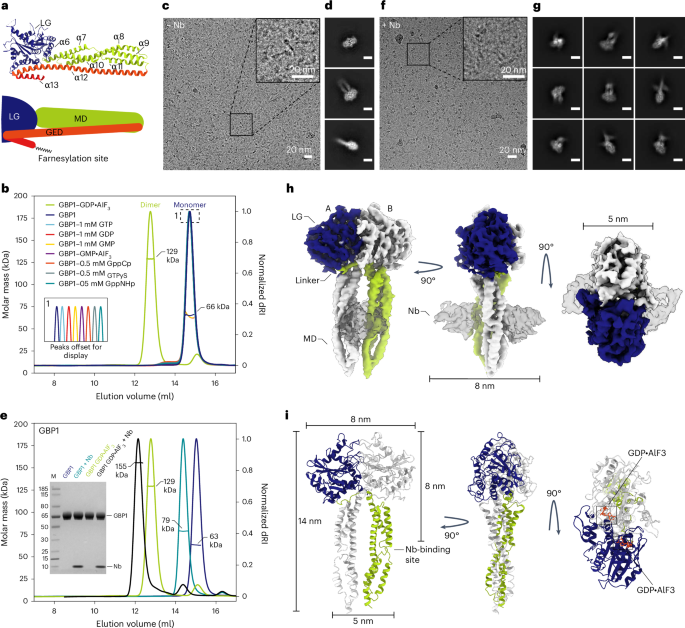
{"title":"Structural basis of antimicrobial membrane coat assembly by human GBP1","authors":"Tanja Kuhm, Clémence Taisne, Cecilia de Agrela Pinto, Luca Gross, Evdokia A. Giannopoulou, Stefan T. Huber, Els Pardon, Jan Steyaert, Sander J. Tans, Arjen J. Jakobi","doi":"10.1038/s41594-024-01400-9","DOIUrl":null,"url":null,"abstract":"Guanylate-binding proteins (GBPs) are interferon-inducible guanosine triphosphate hydrolases (GTPases) mediating host defense against intracellular pathogens. Their antimicrobial activity hinges on their ability to self-associate and coat pathogen-associated compartments or cytosolic bacteria. Coat formation depends on GTPase activity but how nucleotide binding and hydrolysis prime coat formation remains unclear. Here, we report the cryo-electron microscopy structure of the full-length human GBP1 dimer in its guanine nucleotide-bound state and describe the molecular ultrastructure of the GBP1 coat on liposomes and bacterial lipopolysaccharide membranes. Conformational changes of the middle and GTPase effector domains expose the isoprenylated C terminus for membrane association. The α-helical middle domains form a parallel, crossover arrangement essential for coat formation and position the extended effector domain for intercalation into the lipopolysaccharide layer of gram-negative membranes. Nucleotide binding and hydrolysis create oligomeric scaffolds with contractile abilities that promote membrane extrusion and fragmentation. Our data offer a structural and mechanistic framework for understanding GBP1 effector functions in intracellular immunity. Kuhm et al. reveal how human guanylate-binding protein 1 (GBP1) dimers self-associate to coat bacterial pathogens and uncover a guanosine triphosphate hydrolase-dependent membrane-remodeling activity of GBP1 that is crucial for intracellular immunity","PeriodicalId":49141,"journal":{"name":"Nature Structural & Molecular Biology","volume":"32 1","pages":"172-184"},"PeriodicalIF":10.1000,"publicationDate":"2024-10-11","publicationTypes":"Journal Article","fieldsOfStudy":null,"isOpenAccess":false,"openAccessPdf":"https://www.nature.com/articles/s41594-024-01400-9.pdf","citationCount":"0","resultStr":null,"platform":"Semanticscholar","paperid":null,"PeriodicalName":"Nature Structural & Molecular Biology","FirstCategoryId":"99","ListUrlMain":"https://www.nature.com/articles/s41594-024-01400-9","RegionNum":1,"RegionCategory":"生物学","ArticlePicture":[],"TitleCN":null,"AbstractTextCN":null,"PMCID":null,"EPubDate":"","PubModel":"","JCR":"Q1","JCRName":"BIOCHEMISTRY & MOLECULAR BIOLOGY","Score":null,"Total":0}
引用次数: 0
Abstract
Guanylate-binding proteins (GBPs) are interferon-inducible guanosine triphosphate hydrolases (GTPases) mediating host defense against intracellular pathogens. Their antimicrobial activity hinges on their ability to self-associate and coat pathogen-associated compartments or cytosolic bacteria. Coat formation depends on GTPase activity but how nucleotide binding and hydrolysis prime coat formation remains unclear. Here, we report the cryo-electron microscopy structure of the full-length human GBP1 dimer in its guanine nucleotide-bound state and describe the molecular ultrastructure of the GBP1 coat on liposomes and bacterial lipopolysaccharide membranes. Conformational changes of the middle and GTPase effector domains expose the isoprenylated C terminus for membrane association. The α-helical middle domains form a parallel, crossover arrangement essential for coat formation and position the extended effector domain for intercalation into the lipopolysaccharide layer of gram-negative membranes. Nucleotide binding and hydrolysis create oligomeric scaffolds with contractile abilities that promote membrane extrusion and fragmentation. Our data offer a structural and mechanistic framework for understanding GBP1 effector functions in intracellular immunity. Kuhm et al. reveal how human guanylate-binding protein 1 (GBP1) dimers self-associate to coat bacterial pathogens and uncover a guanosine triphosphate hydrolase-dependent membrane-remodeling activity of GBP1 that is crucial for intracellular immunity
期刊介绍:
Nature Structural & Molecular Biology is a comprehensive platform that combines structural and molecular research. Our journal focuses on exploring the functional and mechanistic aspects of biological processes, emphasizing how molecular components collaborate to achieve a particular function. While structural data can shed light on these insights, our publication does not require them as a prerequisite.


 分享
分享
 求助内容:
求助内容: 应助结果提醒方式:
 扫码关注我们
