{"title":"Time-course remodeling and pathology intervention of α-synuclein amyloid fibril by heparin and heparin-like oligosaccharides","authors":"Youqi Tao, Peng Xu, Shenqing Zhang, Wei Shangguan, Guang Yang, Kaien Liu, Xiang Li, Yunpeng Sun, Qinyue Zhao, Dan Li, Biao Yu, Cong Liu","doi":"10.1038/s41594-024-01407-2","DOIUrl":null,"url":null,"abstract":"Amyloid fibrils represent a pathological state of protein polymer that is closely associated with various neurodegenerative diseases. Polysaccharides have a prominent role in recognizing amyloid fibrils and mediating their pathogenicity. However, the mechanism underlying the amyloid–polysaccharide interaction remains elusive. We also do not know its impact on the structure and pathology of formed fibrils. Here, we used cryo-electron microscopy to analyze the atomic structures of mature α-synuclein (α-syn) fibrils upon binding with polymeric heparin and heparin-like oligosaccharides. The fibril structure, including the helical twist and conformation of α-syn, changed over time upon the binding of heparin but not oligosaccharides. The sulfation pattern and numbers of saccharide units are important for the binding. Similarly, negatively charged biopolymers typically interact with amyloid fibrils, including tau and various α-syn polymorphs, leading to alterations in their conformation. Moreover, we show that heparin-like oligosaccharides can not only block neuronal uptake and propagation of formed α-syn fibrils but also inhibit α-syn fibrillation. This work demonstrates a distinctive activity of heparin and biopolymers in remodeling amyloid fibrils and suggests the pharmaceutical potential of heparin-like oligosaccharides. Here, the authors reveal progressive conformational changes of α-synuclein fibrils upon binding with polysaccharide heparin, suggesting that biopolymers have a crucial role in remodeling the structures and pathological activities of amyloid fibrils.","PeriodicalId":49141,"journal":{"name":"Nature Structural & Molecular Biology","volume":"32 2","pages":"369-380"},"PeriodicalIF":10.1000,"publicationDate":"2024-10-17","publicationTypes":"Journal Article","fieldsOfStudy":null,"isOpenAccess":false,"openAccessPdf":"","citationCount":"0","resultStr":null,"platform":"Semanticscholar","paperid":null,"PeriodicalName":"Nature Structural & Molecular Biology","FirstCategoryId":"99","ListUrlMain":"https://www.nature.com/articles/s41594-024-01407-2","RegionNum":1,"RegionCategory":"生物学","ArticlePicture":[],"TitleCN":null,"AbstractTextCN":null,"PMCID":null,"EPubDate":"","PubModel":"","JCR":"Q1","JCRName":"BIOCHEMISTRY & MOLECULAR BIOLOGY","Score":null,"Total":0}
引用次数: 0
Abstract
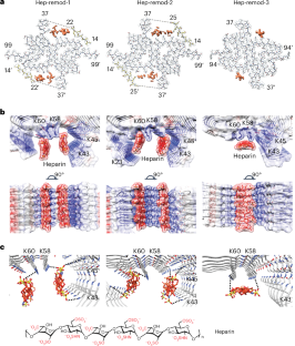
Amyloid fibrils represent a pathological state of protein polymer that is closely associated with various neurodegenerative diseases. Polysaccharides have a prominent role in recognizing amyloid fibrils and mediating their pathogenicity. However, the mechanism underlying the amyloid–polysaccharide interaction remains elusive. We also do not know its impact on the structure and pathology of formed fibrils. Here, we used cryo-electron microscopy to analyze the atomic structures of mature α-synuclein (α-syn) fibrils upon binding with polymeric heparin and heparin-like oligosaccharides. The fibril structure, including the helical twist and conformation of α-syn, changed over time upon the binding of heparin but not oligosaccharides. The sulfation pattern and numbers of saccharide units are important for the binding. Similarly, negatively charged biopolymers typically interact with amyloid fibrils, including tau and various α-syn polymorphs, leading to alterations in their conformation. Moreover, we show that heparin-like oligosaccharides can not only block neuronal uptake and propagation of formed α-syn fibrils but also inhibit α-syn fibrillation. This work demonstrates a distinctive activity of heparin and biopolymers in remodeling amyloid fibrils and suggests the pharmaceutical potential of heparin-like oligosaccharides. Here, the authors reveal progressive conformational changes of α-synuclein fibrils upon binding with polysaccharide heparin, suggesting that biopolymers have a crucial role in remodeling the structures and pathological activities of amyloid fibrils.
期刊介绍:
Nature Structural & Molecular Biology is a comprehensive platform that combines structural and molecular research. Our journal focuses on exploring the functional and mechanistic aspects of biological processes, emphasizing how molecular components collaborate to achieve a particular function. While structural data can shed light on these insights, our publication does not require them as a prerequisite.


 分享
分享
 求助内容:
求助内容: 应助结果提醒方式:
 扫码关注我们
