Jabi Elijah Shriki, Ashley Elizabeth Prosper, Jerold Shinbane, Patrick M Colletti
{"title":"Frequency of myocardial infarcts on conventional, non-gated CT: An often-overlooked entity.","authors":"Jabi Elijah Shriki, Ashley Elizabeth Prosper, Jerold Shinbane, Patrick M Colletti","doi":"10.25259/JCIS_114_2024","DOIUrl":null,"url":null,"abstract":"<p><strong>Objectives: </strong>The objective of this study was to determine how often myocardial infarctions are retrospectively visible on conventional, non-gated, non-cardiac computed tomography (CT) scans. Our goal was to evaluate a cohort of patients with myocardial infarctions visible on cardiac magnetic resonance imaging (MRI) to determine how often the area of infarction was retrospectively visible by preceding, conventional CT. We also sought to evaluate how often the diagnosis of myocardial infarction was reported at the time of initial study review.</p><p><strong>Material and methods: </strong>The Institutional Review Board approval was obtained for the creation and retrospective analysis of a database of patients undergoing cardiac MRI. We started with a cohort of 252 patients who had undergone cardiac MRI at our institution, over a 4-year period. We identified 160 patients who had a myocardial infarct visible on MRI.</p><p><strong>Results: </strong>Of the 160 patients who had been identified as having an infarct on cardiac MRI, 54 patients had undergone a recent (within 30 days) conventional CT scan, usually done for non-cardiac indications. In addition to the review of reports, non-cardiac CT scans were also evaluated retrospectively by two experienced, cardiothoracic imaging physicians, including a radiologist and a cardiologist. In 26 of these patients (48.1%), an infarct was visible on the CT images. In 12 of these 26 cases (46.1%), the infarct was noted in the initial report. In the remaining 14 of these 26 cases (53.8%), the infarct was unrecognized at the time of initial study interpretation.</p><p><strong>Conclusion: </strong>Our retrospective analysis demonstrates that myocardial infarctions may be frequently observed on non-gated, non-cardiac CT scans but may be underrecognized and under-reported.</p>","PeriodicalId":15512,"journal":{"name":"Journal of Clinical Imaging Science","volume":"14 ","pages":"45"},"PeriodicalIF":1.3000,"publicationDate":"2024-11-21","publicationTypes":"Journal Article","fieldsOfStudy":null,"isOpenAccess":false,"openAccessPdf":"https://www.ncbi.nlm.nih.gov/pmc/articles/PMC11618727/pdf/","citationCount":"0","resultStr":null,"platform":"Semanticscholar","paperid":null,"PeriodicalName":"Journal of Clinical Imaging Science","FirstCategoryId":"1085","ListUrlMain":"https://doi.org/10.25259/JCIS_114_2024","RegionNum":0,"RegionCategory":null,"ArticlePicture":[],"TitleCN":null,"AbstractTextCN":null,"PMCID":null,"EPubDate":"2024/1/1 0:00:00","PubModel":"eCollection","JCR":"Q3","JCRName":"RADIOLOGY, NUCLEAR MEDICINE & MEDICAL IMAGING","Score":null,"Total":0}
引用次数: 0
Abstract
Objectives: The objective of this study was to determine how often myocardial infarctions are retrospectively visible on conventional, non-gated, non-cardiac computed tomography (CT) scans. Our goal was to evaluate a cohort of patients with myocardial infarctions visible on cardiac magnetic resonance imaging (MRI) to determine how often the area of infarction was retrospectively visible by preceding, conventional CT. We also sought to evaluate how often the diagnosis of myocardial infarction was reported at the time of initial study review.
Material and methods: The Institutional Review Board approval was obtained for the creation and retrospective analysis of a database of patients undergoing cardiac MRI. We started with a cohort of 252 patients who had undergone cardiac MRI at our institution, over a 4-year period. We identified 160 patients who had a myocardial infarct visible on MRI.
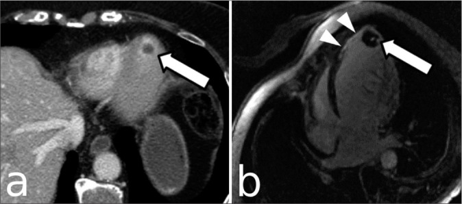
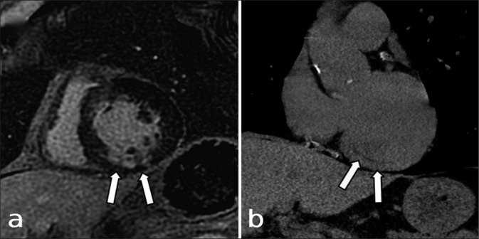
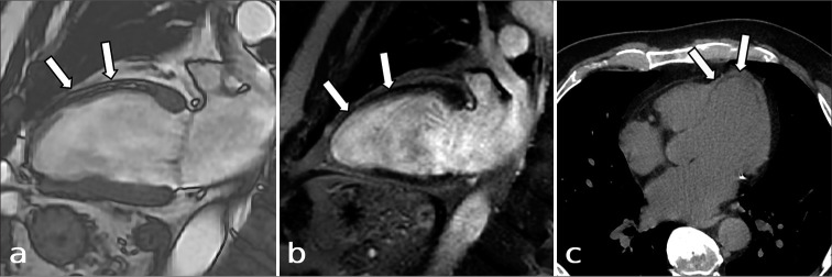
Results: Of the 160 patients who had been identified as having an infarct on cardiac MRI, 54 patients had undergone a recent (within 30 days) conventional CT scan, usually done for non-cardiac indications. In addition to the review of reports, non-cardiac CT scans were also evaluated retrospectively by two experienced, cardiothoracic imaging physicians, including a radiologist and a cardiologist. In 26 of these patients (48.1%), an infarct was visible on the CT images. In 12 of these 26 cases (46.1%), the infarct was noted in the initial report. In the remaining 14 of these 26 cases (53.8%), the infarct was unrecognized at the time of initial study interpretation.
Conclusion: Our retrospective analysis demonstrates that myocardial infarctions may be frequently observed on non-gated, non-cardiac CT scans but may be underrecognized and under-reported.
期刊介绍:
The Journal of Clinical Imaging Science (JCIS) is an open access peer-reviewed journal committed to publishing high-quality articles in the field of Imaging Science. The journal aims to present Imaging Science and relevant clinical information in an understandable and useful format. The journal is owned and published by the Scientific Scholar. Audience Our audience includes Radiologists, Researchers, Clinicians, medical professionals and students. Review process JCIS has a highly rigorous peer-review process that makes sure that manuscripts are scientifically accurate, relevant, novel and important. Authors disclose all conflicts, affiliations and financial associations such that the published content is not biased.


 分享
分享
 求助内容:
求助内容: 应助结果提醒方式:
 扫码关注我们
