{"title":"Non-Invasive Anatomical Level Cerebrovascular Imaging of Mice Using Diffusion Model-Enhanced Fluorescence Imaging","authors":"Huijie Wu, Yufang He, Zeyu Liu, Peng Zhang, Fan Song, Chenbin Ma, Ruxin Cai, Guanglei Zhang","doi":"10.1002/lpor.202401193","DOIUrl":null,"url":null,"abstract":"<p>In vivo fluorescence imaging, particularly indocyanine green (ICG)-based imaging, has gained traction for cerebrovascular imaging due to its real-time dynamics, free radiation, and accessibility. However, the presence of the scalp and skull significantly hampers imaging quality, often necessitating invasive procedures or biotoxic probes to achieve adequate depth and resolution. This limitation restricts the broader clinical/preclinical application of fluorescence imaging techniques. To address this, a novel approach is introduced that utilizes deep learning techniques to enhance ICG-based imaging, achieving high-resolution cerebrovascular imaging without invasive methods or biotoxic probes. By leveraging diffusion models, a connection between trans-scalp (TS) and trans-cranial (TC) ICG fluorescence images are establish in the latent space. This allows the transformation of blurred TS images into high-resolution images resembling TC images. Notably, intracerebral vascular structures and microvascular branches are unambiguously observed, achieving an anatomical resolution of 20.1 µm and a 1.7-fold improvement in spatial resolution. Validation also in a mouse model of middle cerebral artery occlusion demonstrates effective and sensitive identification of ischemic stroke sites. This advancement offers a non-invasive, cost-efficient alternative to current expensive imaging methods, paving the way for more advanced fluorescence imaging techniques.</p>","PeriodicalId":204,"journal":{"name":"Laser & Photonics Reviews","volume":"19 7","pages":""},"PeriodicalIF":10.0000,"publicationDate":"2025-01-10","publicationTypes":"Journal Article","fieldsOfStudy":null,"isOpenAccess":false,"openAccessPdf":"","citationCount":"0","resultStr":null,"platform":"Semanticscholar","paperid":null,"PeriodicalName":"Laser & Photonics Reviews","FirstCategoryId":"101","ListUrlMain":"https://onlinelibrary.wiley.com/doi/10.1002/lpor.202401193","RegionNum":1,"RegionCategory":"物理与天体物理","ArticlePicture":[],"TitleCN":null,"AbstractTextCN":null,"PMCID":null,"EPubDate":"","PubModel":"","JCR":"Q1","JCRName":"OPTICS","Score":null,"Total":0}
引用次数: 0
Abstract
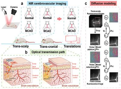
In vivo fluorescence imaging, particularly indocyanine green (ICG)-based imaging, has gained traction for cerebrovascular imaging due to its real-time dynamics, free radiation, and accessibility. However, the presence of the scalp and skull significantly hampers imaging quality, often necessitating invasive procedures or biotoxic probes to achieve adequate depth and resolution. This limitation restricts the broader clinical/preclinical application of fluorescence imaging techniques. To address this, a novel approach is introduced that utilizes deep learning techniques to enhance ICG-based imaging, achieving high-resolution cerebrovascular imaging without invasive methods or biotoxic probes. By leveraging diffusion models, a connection between trans-scalp (TS) and trans-cranial (TC) ICG fluorescence images are establish in the latent space. This allows the transformation of blurred TS images into high-resolution images resembling TC images. Notably, intracerebral vascular structures and microvascular branches are unambiguously observed, achieving an anatomical resolution of 20.1 µm and a 1.7-fold improvement in spatial resolution. Validation also in a mouse model of middle cerebral artery occlusion demonstrates effective and sensitive identification of ischemic stroke sites. This advancement offers a non-invasive, cost-efficient alternative to current expensive imaging methods, paving the way for more advanced fluorescence imaging techniques.
期刊介绍:
Laser & Photonics Reviews is a reputable journal that publishes high-quality Reviews, original Research Articles, and Perspectives in the field of photonics and optics. It covers both theoretical and experimental aspects, including recent groundbreaking research, specific advancements, and innovative applications.
As evidence of its impact and recognition, Laser & Photonics Reviews boasts a remarkable 2022 Impact Factor of 11.0, according to the Journal Citation Reports from Clarivate Analytics (2023). Moreover, it holds impressive rankings in the InCites Journal Citation Reports: in 2021, it was ranked 6th out of 101 in the field of Optics, 15th out of 161 in Applied Physics, and 12th out of 69 in Condensed Matter Physics.
The journal uses the ISSN numbers 1863-8880 for print and 1863-8899 for online publications.


 分享
分享
 求助内容:
求助内容: 应助结果提醒方式:
 扫码关注我们
