Mark Reed, Christopher Miller, Cortney Connor, Jason S Chang, Forshing Lui
{"title":"Fat droplets in the cerebrospinal fluid (CSF) spaces of the brain.","authors":"Mark Reed, Christopher Miller, Cortney Connor, Jason S Chang, Forshing Lui","doi":"10.3934/Neuroscience.2024029","DOIUrl":null,"url":null,"abstract":"<p><p>It is rare to find free floating fat droplets in the cerebral spinal fluid (CSF) spaces of the brain. When fat droplets are seen in the CSF spaces, the most common cause is the rupture of a dermoid cyst. Dermoid cysts are congenital inclusion cysts that form during the neural tube closure between the third and fifth weeks of embryogenesis. In this case report, we describe a case of a 74-year-old, right-handed female who presented with an acute onset of visual disturbances and left-hand numbness. Computed tomography (CT) and magnetic resonance imaging (MRI) of the head revealed hypodense \"lesions\" in the lateral ventricles and basal cisterns. The CT Hounsfield unit was between -41 to -83 Hounsfield Units, which is compatible with fat rather than air. The T1 weighted and FLAIR MRI showed hyperintense lesions \"floating\" on top of the CSF in the lateral ventricles, which is typical for fat droplets, presumably caused by a ruptured dermoid cyst. This case emphasizes the importance of analyzing Hounsfield Units to distinguish lesions by density, where fat ranges from -50 to -150 Hounsfield Units and air is -1000 Hounsfield Units. Pneumocephalus is the presence of air in the epidural, subdural, or subarachnoid space and can cause confusion, nausea, seizures and focal neurological symptoms. A careful analysis of the neuroimaging findings in the CT with or without MRI is important in making a correct diagnosis of a ruptured dermoid cyst versus pneumocephalus.</p>","PeriodicalId":7732,"journal":{"name":"AIMS Neuroscience","volume":"11 4","pages":"484-489"},"PeriodicalIF":2.7000,"publicationDate":"2024-11-27","publicationTypes":"Journal Article","fieldsOfStudy":null,"isOpenAccess":false,"openAccessPdf":"https://www.ncbi.nlm.nih.gov/pmc/articles/PMC11712232/pdf/","citationCount":"0","resultStr":null,"platform":"Semanticscholar","paperid":null,"PeriodicalName":"AIMS Neuroscience","FirstCategoryId":"1085","ListUrlMain":"https://doi.org/10.3934/Neuroscience.2024029","RegionNum":0,"RegionCategory":null,"ArticlePicture":[],"TitleCN":null,"AbstractTextCN":null,"PMCID":null,"EPubDate":"2024/1/1 0:00:00","PubModel":"eCollection","JCR":"Q2","JCRName":"NEUROSCIENCES","Score":null,"Total":0}
引用次数: 0
Abstract
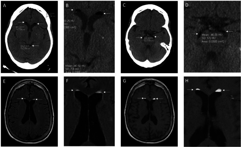
It is rare to find free floating fat droplets in the cerebral spinal fluid (CSF) spaces of the brain. When fat droplets are seen in the CSF spaces, the most common cause is the rupture of a dermoid cyst. Dermoid cysts are congenital inclusion cysts that form during the neural tube closure between the third and fifth weeks of embryogenesis. In this case report, we describe a case of a 74-year-old, right-handed female who presented with an acute onset of visual disturbances and left-hand numbness. Computed tomography (CT) and magnetic resonance imaging (MRI) of the head revealed hypodense "lesions" in the lateral ventricles and basal cisterns. The CT Hounsfield unit was between -41 to -83 Hounsfield Units, which is compatible with fat rather than air. The T1 weighted and FLAIR MRI showed hyperintense lesions "floating" on top of the CSF in the lateral ventricles, which is typical for fat droplets, presumably caused by a ruptured dermoid cyst. This case emphasizes the importance of analyzing Hounsfield Units to distinguish lesions by density, where fat ranges from -50 to -150 Hounsfield Units and air is -1000 Hounsfield Units. Pneumocephalus is the presence of air in the epidural, subdural, or subarachnoid space and can cause confusion, nausea, seizures and focal neurological symptoms. A careful analysis of the neuroimaging findings in the CT with or without MRI is important in making a correct diagnosis of a ruptured dermoid cyst versus pneumocephalus.
期刊介绍:
AIMS Neuroscience is an international Open Access journal devoted to publishing peer-reviewed, high quality, original papers from all areas in the field of neuroscience. The primary focus is to provide a forum in which to expedite the speed with which theoretical neuroscience progresses toward generating testable hypotheses. In the presence of current and developing technology that offers unprecedented access to functions of the nervous system at all levels, the journal is designed to serve the role of providing the widest variety of the best theoretical views leading to suggested studies. Single blind peer review is provided for all articles and commentaries.


 分享
分享
 求助内容:
求助内容: 应助结果提醒方式:
 扫码关注我们
