{"title":"Multidimensional structural analyses revealed a correlation between thalamic atrophy and white matter degeneration in idiopathic dystonia.","authors":"Jinping Xu, Qinxiu Cheng, Yue Zhang, Yuhan Luo, Linchang Zhong, Huiming Liu, Haoran Zhang, Zhengkun Yang, Jiana Zhang, Zilin Ou, Zhicong Yan, Kangqiang Peng, Gang Liu","doi":"10.1093/braincomms/fcaf026","DOIUrl":null,"url":null,"abstract":"<p><p>Although aberrant changes in grey and white matter are core features of idiopathic dystonia, few studies have explored the correlation between grey and white matter changes in this disease. This study aimed to investigate the coupling correlation between morphological and microstructural alterations in patients with idiopathic dystonia. Structural T1 imaging and diffusion tensor imaging were performed on a relatively large cohort of patients. Multidimensional structural analyses, including voxel-based analyses, voxel-based morphology, fixel-based analyses and surface-based morphometry, were performed to explore these structural alterations. Probabilistic tractography and correlation analyses were employed to examine these relationships. A total of 147 patients with idiopathic dystonia and 137 healthy controls were recruited in this study. There were no significant differences in the cortical morphometry between patients with idiopathic dystonia and healthy controls using voxel- and surface-based morphometry. However, the grey matter volume of the bilateral thalamus, fractional anisotropy in the right anterior corona radiata, right retrolenticular part of the internal capsule and right posterior corona radiata, and the fibre density and cross-section combined in the fibre tract connecting the left ventral posterolateral thalamic nucleus and left area 5 m, were significantly decreased in patients with idiopathic dystonia compared with those in healthy controls. Furthermore, the reduced grey matter volume in the right thalamus not only correlated with the disease duration but also with the reduced fractional anisotropy in the right posterior corona radiata and decreased the fibre density and cross-section combined in the fibre tract connecting the left ventral posterolateral thalamic nucleus and the left area 5 m in patients with idiopathic dystonia. These findings suggest that the thalamus is structurally impaired in idiopathic dystonia and that microstructural disruption in thalamocortical projections occurs secondary to thalamic atrophy.</p>","PeriodicalId":93915,"journal":{"name":"Brain communications","volume":"7 1","pages":"fcaf026"},"PeriodicalIF":4.5000,"publicationDate":"2025-01-20","publicationTypes":"Journal Article","fieldsOfStudy":null,"isOpenAccess":false,"openAccessPdf":"https://www.ncbi.nlm.nih.gov/pmc/articles/PMC11775609/pdf/","citationCount":"0","resultStr":null,"platform":"Semanticscholar","paperid":null,"PeriodicalName":"Brain communications","FirstCategoryId":"1085","ListUrlMain":"https://doi.org/10.1093/braincomms/fcaf026","RegionNum":0,"RegionCategory":null,"ArticlePicture":[],"TitleCN":null,"AbstractTextCN":null,"PMCID":null,"EPubDate":"2025/1/1 0:00:00","PubModel":"eCollection","JCR":"Q1","JCRName":"CLINICAL NEUROLOGY","Score":null,"Total":0}
引用次数: 0
Abstract
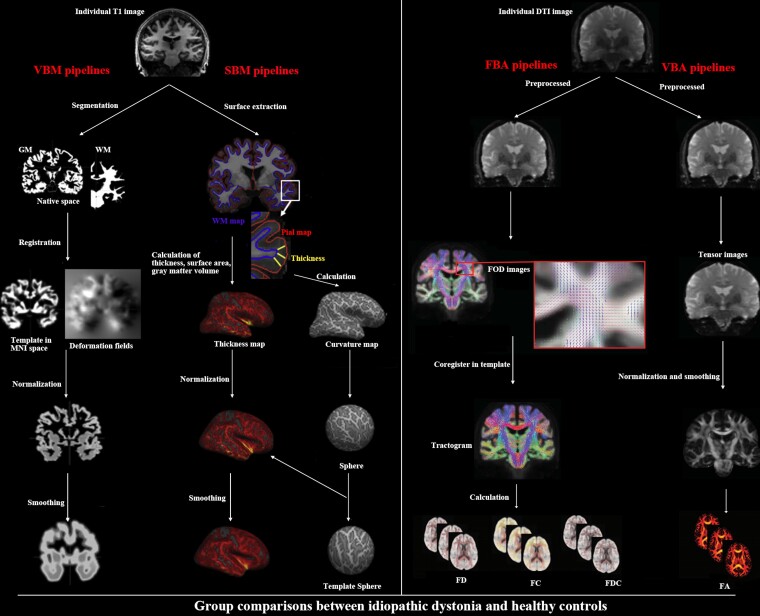
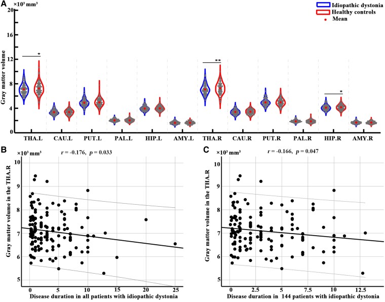
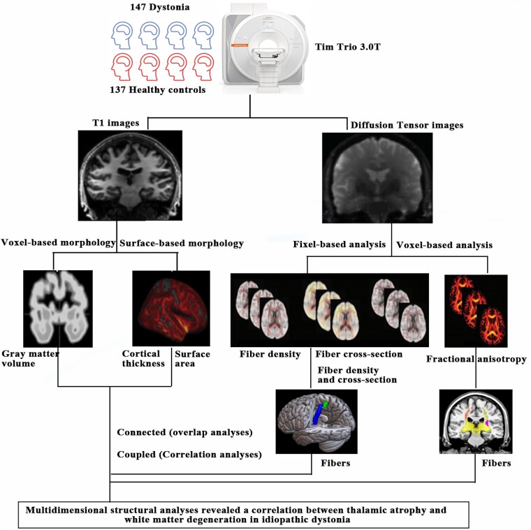
Although aberrant changes in grey and white matter are core features of idiopathic dystonia, few studies have explored the correlation between grey and white matter changes in this disease. This study aimed to investigate the coupling correlation between morphological and microstructural alterations in patients with idiopathic dystonia. Structural T1 imaging and diffusion tensor imaging were performed on a relatively large cohort of patients. Multidimensional structural analyses, including voxel-based analyses, voxel-based morphology, fixel-based analyses and surface-based morphometry, were performed to explore these structural alterations. Probabilistic tractography and correlation analyses were employed to examine these relationships. A total of 147 patients with idiopathic dystonia and 137 healthy controls were recruited in this study. There were no significant differences in the cortical morphometry between patients with idiopathic dystonia and healthy controls using voxel- and surface-based morphometry. However, the grey matter volume of the bilateral thalamus, fractional anisotropy in the right anterior corona radiata, right retrolenticular part of the internal capsule and right posterior corona radiata, and the fibre density and cross-section combined in the fibre tract connecting the left ventral posterolateral thalamic nucleus and left area 5 m, were significantly decreased in patients with idiopathic dystonia compared with those in healthy controls. Furthermore, the reduced grey matter volume in the right thalamus not only correlated with the disease duration but also with the reduced fractional anisotropy in the right posterior corona radiata and decreased the fibre density and cross-section combined in the fibre tract connecting the left ventral posterolateral thalamic nucleus and the left area 5 m in patients with idiopathic dystonia. These findings suggest that the thalamus is structurally impaired in idiopathic dystonia and that microstructural disruption in thalamocortical projections occurs secondary to thalamic atrophy.


 分享
分享

 求助内容:
求助内容: 应助结果提醒方式:
 扫码关注我们
