Hyunsuk Yoo, Hee Eun Moon, Soojin Kim, Da Hee Kim, Young Hun Choi, Jeong-Eun Cheon, Joon Sung Lee, Seunghyun Lee
{"title":"Evaluation of Image Quality and Scan Time Efficiency in Accelerated 3D T1-Weighted Pediatric Brain MRI Using Deep Learning-Based Reconstruction.","authors":"Hyunsuk Yoo, Hee Eun Moon, Soojin Kim, Da Hee Kim, Young Hun Choi, Jeong-Eun Cheon, Joon Sung Lee, Seunghyun Lee","doi":"10.3348/kjr.2024.0701","DOIUrl":null,"url":null,"abstract":"<p><strong>Objective: </strong>This study evaluated the effect of an accelerated three-dimensional (3D) T1-weighted pediatric brain MRI protocol using a deep learning (DL)-based reconstruction algorithm on scan time and image quality.</p><p><strong>Materials and methods: </strong>This retrospective study included 46 pediatric patients who underwent conventional and accelerated, pre- and post-contrast, 3D T1-weighted brain MRI using a 3T scanner (SIGNA Premier; GE HealthCare) at a single tertiary referral center between March 1, 2023, and April 30, 2023. Conventional scans were reconstructed using intensity Filter A (Conv), whereas accelerated scans were reconstructed using intensity Filter A (Fast_A) and a DL-based algorithm (Fast_DL). Image quality was assessed quantitatively based on the coefficient of variation, relative contrast, apparent signal-to-noise ratio (aSNR), and apparent contrast-to-noise ratio (aCNR) and qualitatively according to radiologists' ratings of overall image quality, artifacts, noisiness, gray-white matter differentiation, and lesion conspicuity.</p><p><strong>Results: </strong>The acquisition times for the pre- and post-contrast scans were 191 and 135 seconds, respectively, for the conventional scan. With the accelerated protocol, these were reduced to 135 and 80 seconds, achieving time reductions of 29.3% and 40.7%, respectively. DL-based reconstruction significantly reduced the coefficient of variation, improved the aSNR, aCNR, and overall image quality, and reduced the number of artifacts compared with the conventional acquisition method (all <i>P</i> < 0.05). However, the lesion conspicuity remained similar between the two protocols.</p><p><strong>Conclusion: </strong>Utilizing a DL-based reconstruction algorithm in accelerated 3D T1-weighted pediatric brain MRI can significantly shorten the acquisition time, enhance image quality, and reduce artifacts, making it a viable option for pediatric imaging.</p>","PeriodicalId":17881,"journal":{"name":"Korean Journal of Radiology","volume":"26 2","pages":"180-192"},"PeriodicalIF":5.3000,"publicationDate":"2025-02-01","publicationTypes":"Journal Article","fieldsOfStudy":null,"isOpenAccess":false,"openAccessPdf":"https://www.ncbi.nlm.nih.gov/pmc/articles/PMC11794287/pdf/","citationCount":"0","resultStr":null,"platform":"Semanticscholar","paperid":null,"PeriodicalName":"Korean Journal of Radiology","FirstCategoryId":"3","ListUrlMain":"https://doi.org/10.3348/kjr.2024.0701","RegionNum":2,"RegionCategory":"医学","ArticlePicture":[],"TitleCN":null,"AbstractTextCN":null,"PMCID":null,"EPubDate":"","PubModel":"","JCR":"Q1","JCRName":"RADIOLOGY, NUCLEAR MEDICINE & MEDICAL IMAGING","Score":null,"Total":0}
引用次数: 0
Abstract
Objective: This study evaluated the effect of an accelerated three-dimensional (3D) T1-weighted pediatric brain MRI protocol using a deep learning (DL)-based reconstruction algorithm on scan time and image quality.
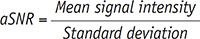
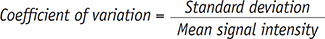
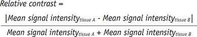
Materials and methods: This retrospective study included 46 pediatric patients who underwent conventional and accelerated, pre- and post-contrast, 3D T1-weighted brain MRI using a 3T scanner (SIGNA Premier; GE HealthCare) at a single tertiary referral center between March 1, 2023, and April 30, 2023. Conventional scans were reconstructed using intensity Filter A (Conv), whereas accelerated scans were reconstructed using intensity Filter A (Fast_A) and a DL-based algorithm (Fast_DL). Image quality was assessed quantitatively based on the coefficient of variation, relative contrast, apparent signal-to-noise ratio (aSNR), and apparent contrast-to-noise ratio (aCNR) and qualitatively according to radiologists' ratings of overall image quality, artifacts, noisiness, gray-white matter differentiation, and lesion conspicuity.
Results: The acquisition times for the pre- and post-contrast scans were 191 and 135 seconds, respectively, for the conventional scan. With the accelerated protocol, these were reduced to 135 and 80 seconds, achieving time reductions of 29.3% and 40.7%, respectively. DL-based reconstruction significantly reduced the coefficient of variation, improved the aSNR, aCNR, and overall image quality, and reduced the number of artifacts compared with the conventional acquisition method (all P < 0.05). However, the lesion conspicuity remained similar between the two protocols.
Conclusion: Utilizing a DL-based reconstruction algorithm in accelerated 3D T1-weighted pediatric brain MRI can significantly shorten the acquisition time, enhance image quality, and reduce artifacts, making it a viable option for pediatric imaging.
期刊介绍:
The inaugural issue of the Korean J Radiol came out in March 2000. Our journal aims to produce and propagate knowledge on radiologic imaging and related sciences.
A unique feature of the articles published in the Journal will be their reflection of global trends in radiology combined with an East-Asian perspective. Geographic differences in disease prevalence will be reflected in the contents of papers, and this will serve to enrich our body of knowledge.
World''s outstanding radiologists from many countries are serving as editorial board of our journal.


 分享
分享
 求助内容:
求助内容: 应助结果提醒方式:
 扫码关注我们
