D. Nascimento , LM Brand , L. Bernardi , JM Vasconcelos , IC Guedes , Figueiredo JAP , F. Visioli , ML Lamers , PV Rados
{"title":"In vitro development of a radicular cyst: A morphological investigation of a spheroid cyst-like model associated with fibroblasts","authors":"D. Nascimento , LM Brand , L. Bernardi , JM Vasconcelos , IC Guedes , Figueiredo JAP , F. Visioli , ML Lamers , PV Rados","doi":"10.1016/j.archoralbio.2025.106186","DOIUrl":null,"url":null,"abstract":"<div><h3>Objective</h3><div>The aim of this study was to improve the <em>in vitro</em> model of tooth radicular cyst previously developed by incorporating stromal components and to describe histologic analysis.</div></div><div><h3>Design</h3><div>A radicular cystogenesis-like 3D model was generated using HaCaT cells (1 × 10<sup>5</sup>) to developing spheroid. After 24 h, spheroids were embedded in non-polymerized collagen in combination with 1 × 10<sup>5</sup> fibroblast cells (HaCaT + 1 × 10<sup>5</sup> hFIB) to mimic stromal microenvironment. Micrographs were taken to evaluate the cystic stability and dispersion area, while histological hematoxylin/eosin staining was used to measure the ratio of epithelial lining area. Analysis was conducted on days 1, 3, and 7 using ImageJ software. Statistical analyses were performed using GraphPad Prism 5 software.</div></div><div><h3>Results</h3><div>The model, with fibroblasts included, preserved the cystic structure and allowed cyst growth, with an increase in both the area and dispersion of the cystic structure throughout the experimental period (p < 0.05). Histological analysis of the cyst model revealed morphological similarities with <em>in vivo</em> tooth radicular cyst biopsies, showing a cystic cavity lined by an epithelial layer, surrounded by collagen and fibroblasts. Additionally, the cavity area increased while the limiting epithelial area decreased. The highest epithelial area-to-total area ratio was observed in day 1 spheroids, while the lowest was found on day 7 (p < 0.05).</div></div><div><h3>Conclusion</h3><div>The incorporation of fibroblasts improved the <em>in vitro</em> cystogenesis model, since it did not interfere with the model’s development and more closely mimicked the <em>in vivo</em> microenvironment.</div></div>","PeriodicalId":8288,"journal":{"name":"Archives of oral biology","volume":"172 ","pages":"Article 106186"},"PeriodicalIF":2.1000,"publicationDate":"2025-04-01","publicationTypes":"Journal Article","fieldsOfStudy":null,"isOpenAccess":false,"openAccessPdf":"","citationCount":"0","resultStr":null,"platform":"Semanticscholar","paperid":null,"PeriodicalName":"Archives of oral biology","FirstCategoryId":"3","ListUrlMain":"https://www.sciencedirect.com/science/article/pii/S0003996925000147","RegionNum":4,"RegionCategory":"医学","ArticlePicture":[],"TitleCN":null,"AbstractTextCN":null,"PMCID":null,"EPubDate":"2025/2/1 0:00:00","PubModel":"Epub","JCR":"Q2","JCRName":"DENTISTRY, ORAL SURGERY & MEDICINE","Score":null,"Total":0}
引用次数: 0
Abstract
Objective
The aim of this study was to improve the in vitro model of tooth radicular cyst previously developed by incorporating stromal components and to describe histologic analysis.
Design
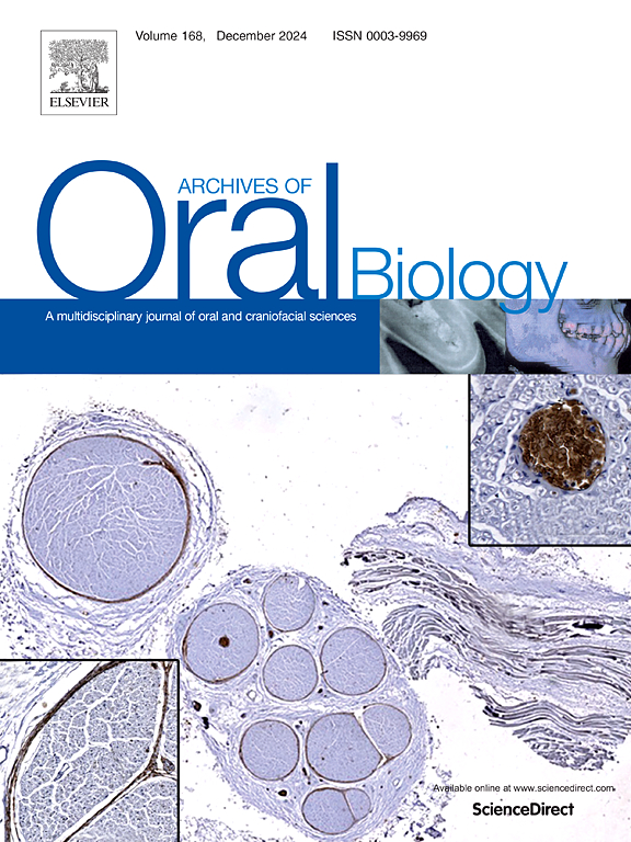
A radicular cystogenesis-like 3D model was generated using HaCaT cells (1 × 10⁵) to developing spheroid. After 24 h, spheroids were embedded in non-polymerized collagen in combination with 1 × 10⁵ fibroblast cells (HaCaT + 1 × 10⁵ hFIB) to mimic stromal microenvironment. Micrographs were taken to evaluate the cystic stability and dispersion area, while histological hematoxylin/eosin staining was used to measure the ratio of epithelial lining area. Analysis was conducted on days 1, 3, and 7 using ImageJ software. Statistical analyses were performed using GraphPad Prism 5 software.
Results
The model, with fibroblasts included, preserved the cystic structure and allowed cyst growth, with an increase in both the area and dispersion of the cystic structure throughout the experimental period (p < 0.05). Histological analysis of the cyst model revealed morphological similarities with in vivo tooth radicular cyst biopsies, showing a cystic cavity lined by an epithelial layer, surrounded by collagen and fibroblasts. Additionally, the cavity area increased while the limiting epithelial area decreased. The highest epithelial area-to-total area ratio was observed in day 1 spheroids, while the lowest was found on day 7 (p < 0.05).
Conclusion
The incorporation of fibroblasts improved the in vitro cystogenesis model, since it did not interfere with the model’s development and more closely mimicked the in vivo microenvironment.
期刊介绍:
Archives of Oral Biology is an international journal which aims to publish papers of the highest scientific quality in the oral and craniofacial sciences. The journal is particularly interested in research which advances knowledge in the mechanisms of craniofacial development and disease, including:
Cell and molecular biology
Molecular genetics
Immunology
Pathogenesis
Cellular microbiology
Embryology
Syndromology
Forensic dentistry


 分享
分享
 求助内容:
求助内容: 应助结果提醒方式:
 扫码关注我们
