Carlos Estrela, Mônica Misaé Endo, Mike Reis Bueno, Bruno Correa Azevedo, Daniel Almeida Decurcio, Lucas Rodrigues Araujo Estrela
{"title":"Application of artifact suppression algorithm of post-processing CBCT software in root canal filling materials.","authors":"Carlos Estrela, Mônica Misaé Endo, Mike Reis Bueno, Bruno Correa Azevedo, Daniel Almeida Decurcio, Lucas Rodrigues Araujo Estrela","doi":"10.1590/1807-3107bor-2025.vol39.011","DOIUrl":null,"url":null,"abstract":"<p><strong>Objectives: </strong>Cone beam computed tomography (CBCT) is an imaging exam used increasingly in various fields of dentistry, and a greater number of endodontists are progressively gaining access to this technology. This study focused on applying an artifact suppression algorithm featured in CBCT software, and designed specifically to address artifacts related to root canal filling materials.</p><p><strong>Method: </strong>The sample consisted of eighty-four mandibular molars, with mesial root canals endodontically treated by using the lateral condensation technique. Four root canal sealers were applied: G1 - Sealapex®, G2 - AH Plus®, G3 - Endofill®, and G4 - Bio-C Sealer. CBCT scans were taken using PreXion 3D Elite®. Initially, the diameter of the root canal filling (in the mesiodistal and buccolingual directions) was measured using a digital micrometer (control). Next, these diameters were reevaluated in the CBCT images using the blooming artifact reduction (BAR) tool of the e-Vol DX software. The Van der Waerden nonparametric analysis of variance was performed, followed by applying the Tukey test to the normalized data. The significance level was set at α = 5%.</p><p><strong>Results: </strong>There were no statistically significant differences (p>0.05) in the measurement of original root canal filling materials obtained by the micrometer versus the e-Vol DX software in the mesiodistal and buccolingual directions.</p><p><strong>Conclusions: </strong>The tested software algorithm effectively suppressed artifacts resulting from obturation materials.</p>","PeriodicalId":9240,"journal":{"name":"Brazilian oral research","volume":"39 ","pages":"e011"},"PeriodicalIF":1.3000,"publicationDate":"2025-02-03","publicationTypes":"Journal Article","fieldsOfStudy":null,"isOpenAccess":false,"openAccessPdf":"https://www.ncbi.nlm.nih.gov/pmc/articles/PMC11790072/pdf/","citationCount":"0","resultStr":null,"platform":"Semanticscholar","paperid":null,"PeriodicalName":"Brazilian oral research","FirstCategoryId":"3","ListUrlMain":"https://doi.org/10.1590/1807-3107bor-2025.vol39.011","RegionNum":4,"RegionCategory":"医学","ArticlePicture":[],"TitleCN":null,"AbstractTextCN":null,"PMCID":null,"EPubDate":"2025/1/1 0:00:00","PubModel":"eCollection","JCR":"Q3","JCRName":"DENTISTRY, ORAL SURGERY & MEDICINE","Score":null,"Total":0}
引用次数: 0
Abstract
Objectives: Cone beam computed tomography (CBCT) is an imaging exam used increasingly in various fields of dentistry, and a greater number of endodontists are progressively gaining access to this technology. This study focused on applying an artifact suppression algorithm featured in CBCT software, and designed specifically to address artifacts related to root canal filling materials.
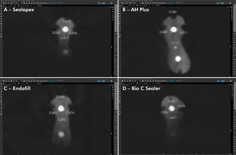
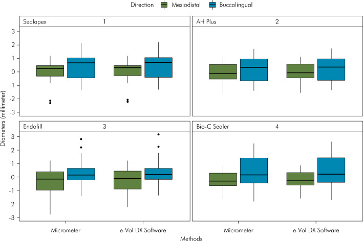
Method: The sample consisted of eighty-four mandibular molars, with mesial root canals endodontically treated by using the lateral condensation technique. Four root canal sealers were applied: G1 - Sealapex®, G2 - AH Plus®, G3 - Endofill®, and G4 - Bio-C Sealer. CBCT scans were taken using PreXion 3D Elite®. Initially, the diameter of the root canal filling (in the mesiodistal and buccolingual directions) was measured using a digital micrometer (control). Next, these diameters were reevaluated in the CBCT images using the blooming artifact reduction (BAR) tool of the e-Vol DX software. The Van der Waerden nonparametric analysis of variance was performed, followed by applying the Tukey test to the normalized data. The significance level was set at α = 5%.
Results: There were no statistically significant differences (p>0.05) in the measurement of original root canal filling materials obtained by the micrometer versus the e-Vol DX software in the mesiodistal and buccolingual directions.
Conclusions: The tested software algorithm effectively suppressed artifacts resulting from obturation materials.


 分享
分享
 求助内容:
求助内容: 应助结果提醒方式:
 扫码关注我们
