Daniel Andrés Ardila Botero, Laura Céspedes Trujillo
{"title":"[Craniopharyngioma with hemorrhagic presentation and visual impairment in a pregnant woman. Case report and literature review].","authors":"Daniel Andrés Ardila Botero, Laura Céspedes Trujillo","doi":"10.18597/rcog.4215","DOIUrl":null,"url":null,"abstract":"<p><strong>Objectives: </strong>To present the case of a pregnant woman diagnosed with craniopharyngioma and to review the literature on the diagnosis, treatment, and maternal-perinatal outcomes of this type of tumor.</p><p><strong>Material and methods: </strong>A 41-year-old multigravida at 23.6 weeks of gestation was admitted to a high-complexity private clinic due to significant bilateral visual acuity reduction and headache. A diagnosis of craniopharyngioma was made, and expectant management was chosen. The patient underwent an uncomplicated cesarean delivery. The literature review included case reports and series on pregnant women diagnosed with craniopharyngioma. Literature was searched on PubMed, EBSCO, and Scopus, along with references from the selected studies. A narrative summary of the findings is provided.</p><p><strong>Results: </strong>Ten case reports were included. All patients presented with visual disturbances, and eight cases had diabetes insipidus. Magnetic resonance imaging (MRI) was used in nine cases, effectively identifying the tumor. Seven patients underwent craniotomy (four during pregnancy, two postpartum, and one post-abortion), while three had transsphenoidal surgery (two during pregnancy and one postpartum). Three cases experienced tumor recurrence, and two had incomplete resection. During the postpartum period, 9 cases had resolution of visual symptoms. In four cases, follow-up ranged from two to six years without evidence of recurrence.</p><p><strong>Conclusions: </strong>In pregnant women with bitemporal hemianopsia, a possible suprasellar tumor should be suspected. MRI of the brain and sella turcica is the diagnostic modality of choice. Further studies are needed to document this condition in pregnancy and its obstetric management in greater detail.</p>","PeriodicalId":101422,"journal":{"name":"Revista colombiana de obstetricia y ginecologia","volume":"75 4","pages":""},"PeriodicalIF":0.0000,"publicationDate":"2024-11-25","publicationTypes":"Journal Article","fieldsOfStudy":null,"isOpenAccess":false,"openAccessPdf":"https://www.ncbi.nlm.nih.gov/pmc/articles/PMC11812093/pdf/","citationCount":"0","resultStr":null,"platform":"Semanticscholar","paperid":null,"PeriodicalName":"Revista colombiana de obstetricia y ginecologia","FirstCategoryId":"1085","ListUrlMain":"https://doi.org/10.18597/rcog.4215","RegionNum":0,"RegionCategory":null,"ArticlePicture":[],"TitleCN":null,"AbstractTextCN":null,"PMCID":null,"EPubDate":"","PubModel":"","JCR":"","JCRName":"","Score":null,"Total":0}
引用次数: 0
Abstract
Objectives: To present the case of a pregnant woman diagnosed with craniopharyngioma and to review the literature on the diagnosis, treatment, and maternal-perinatal outcomes of this type of tumor.
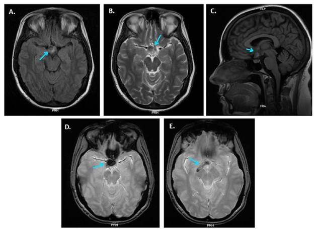
Material and methods: A 41-year-old multigravida at 23.6 weeks of gestation was admitted to a high-complexity private clinic due to significant bilateral visual acuity reduction and headache. A diagnosis of craniopharyngioma was made, and expectant management was chosen. The patient underwent an uncomplicated cesarean delivery. The literature review included case reports and series on pregnant women diagnosed with craniopharyngioma. Literature was searched on PubMed, EBSCO, and Scopus, along with references from the selected studies. A narrative summary of the findings is provided.
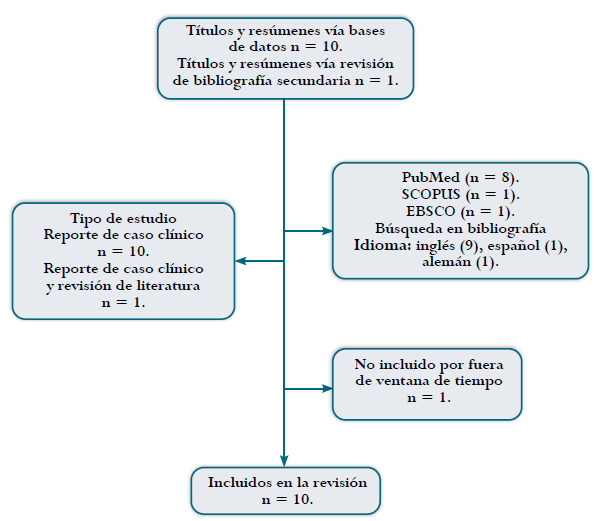
Results: Ten case reports were included. All patients presented with visual disturbances, and eight cases had diabetes insipidus. Magnetic resonance imaging (MRI) was used in nine cases, effectively identifying the tumor. Seven patients underwent craniotomy (four during pregnancy, two postpartum, and one post-abortion), while three had transsphenoidal surgery (two during pregnancy and one postpartum). Three cases experienced tumor recurrence, and two had incomplete resection. During the postpartum period, 9 cases had resolution of visual symptoms. In four cases, follow-up ranged from two to six years without evidence of recurrence.
Conclusions: In pregnant women with bitemporal hemianopsia, a possible suprasellar tumor should be suspected. MRI of the brain and sella turcica is the diagnostic modality of choice. Further studies are needed to document this condition in pregnancy and its obstetric management in greater detail.


 分享
分享

 求助内容:
求助内容: 应助结果提醒方式:
 扫码关注我们
