{"title":"Late gadolinium enhancement in areas with electrically fractionated potentials during sinus rhythm in patients with atrial fibrillation.","authors":"Yuya Suzuki, Kunihiko Kiuchi, Mitsuru Takami, Kimitake Imamura, Jun Sakai, Toshihiro Nakamura, Atsusuke Yatomi, Yusuke Sonoda, Hiroyuki Takahara, Kazutaka Nakasone, Kyoko Yamamoto, Kenichi Tani, Hidehiro Iwai, Yusuke Nakanishi, Mitsuhiko Shoda, Shogo Yonehara, Atushi Murakami, Ken-Ichi Hirata, Koji Fukuzawa","doi":"10.1007/s00380-025-02515-9","DOIUrl":null,"url":null,"abstract":"<p><p>The areas with electrically fractionated potentials (AEFP) during sinus rhythm are related to non-pulmonary vein triggers and may serve as substrates of atrial fibrillation (AF) maintenance. However, the histological properties of these compounds remain unclear. Therefore, we aimed to evaluate the late gadolinium enhancement (LGE) properties of AEFP in patients with AF. We enrolled 15 patients with AF who had undergone LGE magnetic resonance imaging before catheter ablation. AEFP in the left atrium was detected using the HD-Grid and NavX systems after pulmonary vein isolation. We compared LGE properties between AEFP and the surrounding non-fractionated areas (non-AEFP). LGE heterogeneity and density were evaluated through entropy (LGE entropy) and the volume ratio of the enhancement voxel (LGE volume ratio), respectively. Thirty-three AEFP were detected in the left atrium. LGE entropy and LGE volume ratio were significantly higher in AEFP than in non-AEFP [LGE entropy: 6.2 (6.1-6.4) vs. 5.9 (5.8-6.0), p ≤ 0.0001; LGE volume ratio: 23.0% (17.2-29.0%) vs. 10.4% (3.4-20.2%), p ≤ 0.0001]. The atrial voltages did not differ [2.4 (1.3-3.7) vs. 2.5 (1.9-3.1) mV, p = 0.96]. AF recurrence was more significantly found in patients with more than three AEFP than in those without it (log-rank test: p = 0.009). AEFP is likely to be distributed in heterogeneous and moderate LGE areas, regardless of the atrial voltage.</p>","PeriodicalId":12940,"journal":{"name":"Heart and Vessels","volume":" ","pages":"718-725"},"PeriodicalIF":1.5000,"publicationDate":"2025-08-01","publicationTypes":"Journal Article","fieldsOfStudy":null,"isOpenAccess":false,"openAccessPdf":"https://www.ncbi.nlm.nih.gov/pmc/articles/PMC12263775/pdf/","citationCount":"0","resultStr":null,"platform":"Semanticscholar","paperid":null,"PeriodicalName":"Heart and Vessels","FirstCategoryId":"3","ListUrlMain":"https://doi.org/10.1007/s00380-025-02515-9","RegionNum":4,"RegionCategory":"医学","ArticlePicture":[],"TitleCN":null,"AbstractTextCN":null,"PMCID":null,"EPubDate":"2025/2/8 0:00:00","PubModel":"Epub","JCR":"Q3","JCRName":"CARDIAC & CARDIOVASCULAR SYSTEMS","Score":null,"Total":0}
引用次数: 0
Abstract
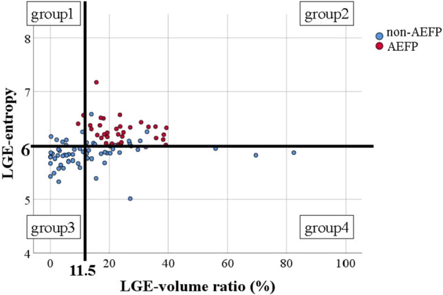
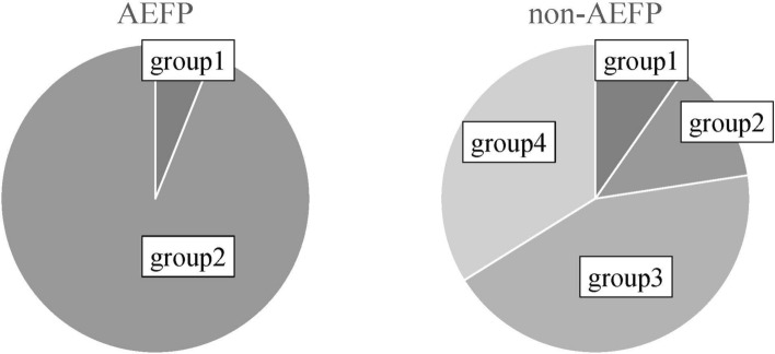
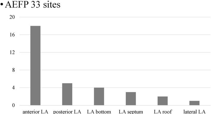
The areas with electrically fractionated potentials (AEFP) during sinus rhythm are related to non-pulmonary vein triggers and may serve as substrates of atrial fibrillation (AF) maintenance. However, the histological properties of these compounds remain unclear. Therefore, we aimed to evaluate the late gadolinium enhancement (LGE) properties of AEFP in patients with AF. We enrolled 15 patients with AF who had undergone LGE magnetic resonance imaging before catheter ablation. AEFP in the left atrium was detected using the HD-Grid and NavX systems after pulmonary vein isolation. We compared LGE properties between AEFP and the surrounding non-fractionated areas (non-AEFP). LGE heterogeneity and density were evaluated through entropy (LGE entropy) and the volume ratio of the enhancement voxel (LGE volume ratio), respectively. Thirty-three AEFP were detected in the left atrium. LGE entropy and LGE volume ratio were significantly higher in AEFP than in non-AEFP [LGE entropy: 6.2 (6.1-6.4) vs. 5.9 (5.8-6.0), p ≤ 0.0001; LGE volume ratio: 23.0% (17.2-29.0%) vs. 10.4% (3.4-20.2%), p ≤ 0.0001]. The atrial voltages did not differ [2.4 (1.3-3.7) vs. 2.5 (1.9-3.1) mV, p = 0.96]. AF recurrence was more significantly found in patients with more than three AEFP than in those without it (log-rank test: p = 0.009). AEFP is likely to be distributed in heterogeneous and moderate LGE areas, regardless of the atrial voltage.
期刊介绍:
Heart and Vessels is an English-language journal that provides a forum of original ideas, excellent methods, and fascinating techniques on cardiovascular disease fields. All papers submitted for publication are evaluated only with regard to scientific quality and relevance to the heart and vessels. Contributions from those engaged in practical medicine, as well as from those involved in basic research, are welcomed.


 分享
分享
 求助内容:
求助内容: 应助结果提醒方式:
 扫码关注我们
