{"title":"Mass Spectrometry as a First-Line Diagnostic Aid for Congenital Disorders of Glycosylation.","authors":"Yoshinao Wada","doi":"10.5702/massspectrometry.A0169","DOIUrl":null,"url":null,"abstract":"<p><p>Congenital disorders of glycosylation (CDG) constitute a group of rare inherited metabolic disorders resulting from mutations in genes involved in the biosynthesis of glycan chains that are covalently attached to proteins or lipids. To date, nearly 200 genes have been identified as responsible for these disorders, with approximately half implicated in N-glycosylation defects. Diagnosis of CDG is primarily achieved through genetic analysis and the identification of glycan abnormalities, referred to as molecular phenotypes. With the increasing use of whole exome and genome sequencing in the investigation of diseases with unknown etiology, the number of cases suspected of CDG is increasing, highlighting the necessity for glycan analysis. Molecular phenotyping in CDG typically targets glycoproteins, with transferrin and apolipoprotein CIII being key representatives of N- and mucin-type O-glycosylation, respectively. Mass spectrometry (MS) provides rapid analysis and yields moderately detailed information, establishing it as a first-line molecular diagnostic tool that complements genetic analysis. Structural anomalies detected by MS can be classified into distinct patterns, which may indicate specific defects within the glycosylation pathway. In cases of CDG types that lack clear molecular phenotypes, characteristic metabolites can often be identified and quantified by MS, further aiding in the diagnostic process. Molecular diagnosis of CDG using MS can be performed with a standard mass spectrometer and a dried blood spot on filter paper, enabling its application in population-based mass screening.</p>","PeriodicalId":18243,"journal":{"name":"Mass spectrometry","volume":"14 1","pages":"A0169"},"PeriodicalIF":0.0000,"publicationDate":"2025-01-01","publicationTypes":"Journal Article","fieldsOfStudy":null,"isOpenAccess":false,"openAccessPdf":"https://www.ncbi.nlm.nih.gov/pmc/articles/PMC11808201/pdf/","citationCount":"0","resultStr":null,"platform":"Semanticscholar","paperid":null,"PeriodicalName":"Mass spectrometry","FirstCategoryId":"1085","ListUrlMain":"https://doi.org/10.5702/massspectrometry.A0169","RegionNum":0,"RegionCategory":null,"ArticlePicture":[],"TitleCN":null,"AbstractTextCN":null,"PMCID":null,"EPubDate":"2025/2/8 0:00:00","PubModel":"Epub","JCR":"Q3","JCRName":"Physics and Astronomy","Score":null,"Total":0}
引用次数: 0
Abstract
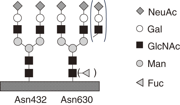
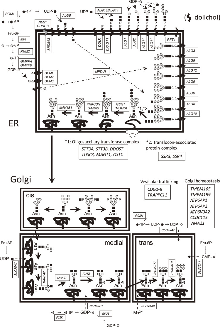
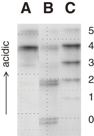
Congenital disorders of glycosylation (CDG) constitute a group of rare inherited metabolic disorders resulting from mutations in genes involved in the biosynthesis of glycan chains that are covalently attached to proteins or lipids. To date, nearly 200 genes have been identified as responsible for these disorders, with approximately half implicated in N-glycosylation defects. Diagnosis of CDG is primarily achieved through genetic analysis and the identification of glycan abnormalities, referred to as molecular phenotypes. With the increasing use of whole exome and genome sequencing in the investigation of diseases with unknown etiology, the number of cases suspected of CDG is increasing, highlighting the necessity for glycan analysis. Molecular phenotyping in CDG typically targets glycoproteins, with transferrin and apolipoprotein CIII being key representatives of N- and mucin-type O-glycosylation, respectively. Mass spectrometry (MS) provides rapid analysis and yields moderately detailed information, establishing it as a first-line molecular diagnostic tool that complements genetic analysis. Structural anomalies detected by MS can be classified into distinct patterns, which may indicate specific defects within the glycosylation pathway. In cases of CDG types that lack clear molecular phenotypes, characteristic metabolites can often be identified and quantified by MS, further aiding in the diagnostic process. Molecular diagnosis of CDG using MS can be performed with a standard mass spectrometer and a dried blood spot on filter paper, enabling its application in population-based mass screening.


 分享
分享
 求助内容:
求助内容: 应助结果提醒方式:
 扫码关注我们
