{"title":"Vimentin intermediate filaments coordinate actin stress fibers and podosomes to determine the extracellular matrix degradation by macrophages","authors":"Xinyi Huang, Zhifang Li, Yuhan Huang, Qian Zhang, Yanqin Cui, Xuemeng Shi, Yaming Jiu","doi":"10.1016/j.devcel.2025.01.016","DOIUrl":null,"url":null,"abstract":"Macrophages possess the capacity to degrade extracellular matrix (ECM), but the specific roles of different cytoskeletal structures in controlling this process are incompletely understood. Here, we report that the inward flow of actin stress fibers delivers endocytosed ECM for lysosomal elimination, replenishing the pool of enzymes for extracellular ECM hydrolysis in actin-rich podosomes. Vimentin deficiency disrupted the balance between stress fibers and podosomes, impairing ECM degradation through integrin CD11b in THP-1 macrophages. In lung adenocarcinoma patient samples, M2-type macrophages exhibit a tighter podosome organization, surrounded by compact vimentin filaments, than M1-type. <em>In vitro</em> experiments verified that the invasion ability of A549 lung carcinoma cells was enhanced when accompanied by wild type, but not vimentin knockout M2-type THP-1, macrophages. Subcutaneous injections of macrophages and tumor cells in nude mice showed that vimentin in macrophages can reduce tumor collagen fibers. Together, our findings provide insights into the cytoskeletal dynamics governing macrophage ECM degradation.","PeriodicalId":11157,"journal":{"name":"Developmental cell","volume":"15 1","pages":""},"PeriodicalIF":8.7000,"publicationDate":"2025-02-13","publicationTypes":"Journal Article","fieldsOfStudy":null,"isOpenAccess":false,"openAccessPdf":"","citationCount":"0","resultStr":null,"platform":"Semanticscholar","paperid":null,"PeriodicalName":"Developmental cell","FirstCategoryId":"99","ListUrlMain":"https://doi.org/10.1016/j.devcel.2025.01.016","RegionNum":1,"RegionCategory":"生物学","ArticlePicture":[],"TitleCN":null,"AbstractTextCN":null,"PMCID":null,"EPubDate":"","PubModel":"","JCR":"Q1","JCRName":"CELL BIOLOGY","Score":null,"Total":0}
引用次数: 0
Abstract
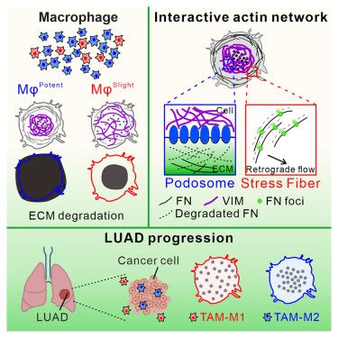
Macrophages possess the capacity to degrade extracellular matrix (ECM), but the specific roles of different cytoskeletal structures in controlling this process are incompletely understood. Here, we report that the inward flow of actin stress fibers delivers endocytosed ECM for lysosomal elimination, replenishing the pool of enzymes for extracellular ECM hydrolysis in actin-rich podosomes. Vimentin deficiency disrupted the balance between stress fibers and podosomes, impairing ECM degradation through integrin CD11b in THP-1 macrophages. In lung adenocarcinoma patient samples, M2-type macrophages exhibit a tighter podosome organization, surrounded by compact vimentin filaments, than M1-type. In vitro experiments verified that the invasion ability of A549 lung carcinoma cells was enhanced when accompanied by wild type, but not vimentin knockout M2-type THP-1, macrophages. Subcutaneous injections of macrophages and tumor cells in nude mice showed that vimentin in macrophages can reduce tumor collagen fibers. Together, our findings provide insights into the cytoskeletal dynamics governing macrophage ECM degradation.
期刊介绍:
Developmental Cell, established in 2001, is a comprehensive journal that explores a wide range of topics in cell and developmental biology. Our publication encompasses work across various disciplines within biology, with a particular emphasis on investigating the intersections between cell biology, developmental biology, and other related fields. Our primary objective is to present research conducted through a cell biological perspective, addressing the essential mechanisms governing cell function, cellular interactions, and responses to the environment. Moreover, we focus on understanding the collective behavior of cells, culminating in the formation of tissues, organs, and whole organisms, while also investigating the consequences of any malfunctions in these intricate processes.


 分享
分享
 求助内容:
求助内容: 应助结果提醒方式:
 扫码关注我们
