{"title":"Evaluation of OCTA-based Parameters in Full-thickness Macular Holes: A Prospective, Comparative, Interventional Study.","authors":"Kalishankar Das, Jit Bhunia, Purban Ganguly, Asim K Ghosh, Debadyuti Chatterjee, Sounak Bepari, Asif Ayub","doi":"10.4103/meajo.meajo_227_22","DOIUrl":null,"url":null,"abstract":"<p><strong>Purpose: </strong>This study aims to correlate optical coherence tomography angiography (OCTA)-based retinal microvasculature changes in cases of full-thickness macular hole (FTMH) before and after vitreoretinal surgery and its relation to patient's visual recovery.</p><p><strong>Methods: </strong>Data of 31 eyes with FTMH were evaluated preoperatively and post-operatively at 6, 12, and 24 weeks for OCTA parameters and compared.</p><p><strong>Results: </strong>93.55% eyes (29 eyes) showed improvement in best-corrected visual acuity at 24 weeks. The mean foveal avascular zone (FAZ) significantly reduced from 0.41 ± 0.13 mm<sup>2</sup> (preoperatively) to 0.25 ± 0.01 mm<sup>2</sup> (postoperatively at 24 weeks). Mean preoperative vessel density (VD) in the superficial vascular plexus (SVP) progressively improved to 24.2% ± 2.2%, 25.2% ± 2.1% and 25.8% ± 2.3% at 6, 12, and 24 weeks respectively from 24.4% ± 2.1% preoperatively (<i>P</i> = 0.0, <i>F</i> = 5.1). The mean VD of foveal region in the SVP significantly improved (<i>P</i> < 0.0, <i>F</i> = 13.9) while that of the parafoveal region did not improve at 24 weeks (<i>P</i> = 0.3, <i>F</i> = 1.2) when compared with its preoperative status. The mean preoperative VD in the deep vascular plexus (DVP) was 20.2% ± 2.6%. It significantly improved at 6, 12, and 24 weeks (20.3% ± 2.4%, 21.8% ± 2.3% and 22.1% ± 2.2%, respectively; <i>P</i> = 0.0, <i>F</i> = 6.9). The mean VD of foveal region and parafoveal region in the DVP showed significant improvement when compared with its preoperative status (<i>P</i> < 0.0, <i>F</i> = 39.3, <i>P</i> < 0.0, <i>F</i> = 13.7).</p><p><strong>Conclusion: </strong>This study showed reduction in mean FAZ area and improvement in mean VD at SVP and DVP in the macula postoperatively. Routine perioperative OCTA-based documentation of macular vascularity in FTMH may throw a light in cases with anatomico-functional postoperative disparities in future.</p>","PeriodicalId":18740,"journal":{"name":"Middle East African Journal of Ophthalmology","volume":"30 4","pages":"229-233"},"PeriodicalIF":0.3000,"publicationDate":"2024-12-02","publicationTypes":"Journal Article","fieldsOfStudy":null,"isOpenAccess":false,"openAccessPdf":"https://www.ncbi.nlm.nih.gov/pmc/articles/PMC11823539/pdf/","citationCount":"0","resultStr":null,"platform":"Semanticscholar","paperid":null,"PeriodicalName":"Middle East African Journal of Ophthalmology","FirstCategoryId":"1085","ListUrlMain":"https://doi.org/10.4103/meajo.meajo_227_22","RegionNum":0,"RegionCategory":null,"ArticlePicture":[],"TitleCN":null,"AbstractTextCN":null,"PMCID":null,"EPubDate":"2023/10/1 0:00:00","PubModel":"eCollection","JCR":"Q4","JCRName":"OPHTHALMOLOGY","Score":null,"Total":0}
引用次数: 0
Abstract
Purpose: This study aims to correlate optical coherence tomography angiography (OCTA)-based retinal microvasculature changes in cases of full-thickness macular hole (FTMH) before and after vitreoretinal surgery and its relation to patient's visual recovery.
Methods: Data of 31 eyes with FTMH were evaluated preoperatively and post-operatively at 6, 12, and 24 weeks for OCTA parameters and compared.
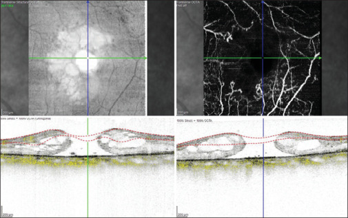
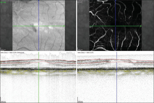
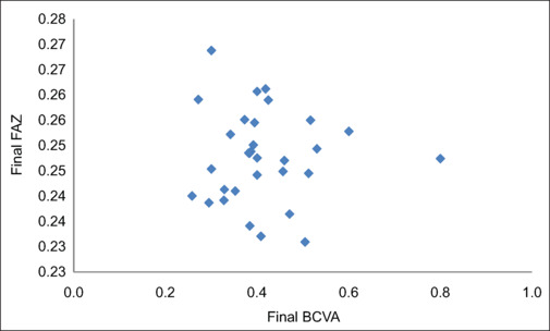
Results: 93.55% eyes (29 eyes) showed improvement in best-corrected visual acuity at 24 weeks. The mean foveal avascular zone (FAZ) significantly reduced from 0.41 ± 0.13 mm2 (preoperatively) to 0.25 ± 0.01 mm2 (postoperatively at 24 weeks). Mean preoperative vessel density (VD) in the superficial vascular plexus (SVP) progressively improved to 24.2% ± 2.2%, 25.2% ± 2.1% and 25.8% ± 2.3% at 6, 12, and 24 weeks respectively from 24.4% ± 2.1% preoperatively (P = 0.0, F = 5.1). The mean VD of foveal region in the SVP significantly improved (P < 0.0, F = 13.9) while that of the parafoveal region did not improve at 24 weeks (P = 0.3, F = 1.2) when compared with its preoperative status. The mean preoperative VD in the deep vascular plexus (DVP) was 20.2% ± 2.6%. It significantly improved at 6, 12, and 24 weeks (20.3% ± 2.4%, 21.8% ± 2.3% and 22.1% ± 2.2%, respectively; P = 0.0, F = 6.9). The mean VD of foveal region and parafoveal region in the DVP showed significant improvement when compared with its preoperative status (P < 0.0, F = 39.3, P < 0.0, F = 13.7).
Conclusion: This study showed reduction in mean FAZ area and improvement in mean VD at SVP and DVP in the macula postoperatively. Routine perioperative OCTA-based documentation of macular vascularity in FTMH may throw a light in cases with anatomico-functional postoperative disparities in future.
目的:研究基于光学相干断层血管造影(OCTA)的全层黄斑孔(FTMH)手术前后视网膜微血管的变化及其与患者视力恢复的关系。方法:对31只FTMH患者术前、术后6、12、24周的OCTA参数进行比较。结果:93.55%眼(29眼)24周最佳矫正视力改善。平均中央凹无血管区(FAZ)从0.41±0.13 mm2(术前)显著降低至0.25±0.01 mm2(术后24周)。术前浅表血管丛(SVP)平均血管密度(VD)在6、12、24周分别由术前的24.4%±2.1%、24.2%±2.2%、25.2%±2.1%和25.8%±2.3%逐步改善(P = 0.0, F = 5.1)。与术前相比,24周SVP的平均中央凹区VD明显改善(P < 0.0, F = 13.9),而中央凹旁区VD无明显改善(P = 0.3, F = 1.2)。术前深血管丛VD (DVP)均值为20.2%±2.6%。6周、12周、24周显著改善(分别为20.3%±2.4%、21.8%±2.3%、22.1%±2.2%);P = 0.0, f = 6.9)。DVP的中央凹区和旁中央凹区平均VD与术前相比有明显改善(P < 0.0, F = 39.3, P < 0.0, F = 13.7)。结论:术后黄斑SVP和DVP区平均FAZ面积减少,平均VD改善。FTMH患者围手术期常规的基于octa的黄斑血管记录可能会对术后解剖功能差异的病例有所帮助。
期刊介绍:
The Middle East African Journal of Ophthalmology (MEAJO), published four times per year in print and online, is an official journal of the Middle East African Council of Ophthalmology (MEACO). It is an international, peer-reviewed journal whose mission includes publication of original research of interest to ophthalmologists in the Middle East and Africa, and to provide readers with high quality educational review articles from world-renown experts. MEAJO, previously known as Middle East Journal of Ophthalmology (MEJO) was founded by Dr Akef El Maghraby in 1993.


 分享
分享
 求助内容:
求助内容: 应助结果提醒方式:
 扫码关注我们
