Dhoha M Alhamad, Asma N AlGhamdi, Batool S AlOtaibi
{"title":"Bilateral Combined Central Retinal Artery and Vein Occlusion in a Child with Purtscher Retinopathy: A Case Report and Review of the Literature.","authors":"Dhoha M Alhamad, Asma N AlGhamdi, Batool S AlOtaibi","doi":"10.4103/meajo.meajo_170_23","DOIUrl":null,"url":null,"abstract":"<p><p>Purtscher retinopathy is a hemorrhagic and vaso-occlusive vasculopathy that results from head trauma. Typically, patients present with sudden onset of painless reduction in visual acuity and a group of retinal findings including retinal hemorrhages, retinal whitening, and optic disc edema. The objectives of the study were to describe the rare occurrence of combined central retinal artery and central vein occlusion in a child with Purtscher retinopathy and to illustrate the course of visual recovery and anatomical changes over 6 months of follow-up. The data were collected from the patient file including circumstances of presentation, visual acuity, anterior segment examination, fundus photography, fundus fluorescein angiography (FFA), B scan ultrasonography (B scan), and optical coherence tomography (OCT). Follow-ups over a 6-month period with repeated imaging were documented. A 6-year-old boy presented with a complaint of bilateral vision loss which occurred 2 days after falling out of high bed. On examination, visual acuity was hand motion bilaterally. External and anterior segments exhibited regular examination, apart from traumatic iritis in both eyes. Posterior segment examination showed intraretinal hemorrhages, retinal whitening bilaterally, and exudative retinal detachment, which was confirmed by OCT and B-scan. FFA of both the eyes showed delayed arterial and venous filling with macular hypoperfusion. Comprehensive systemic workup including brain imaging, hematology, immunology, and uveitis screening was negative. The patient was treated with a high-dose oral steroid. Six months later, his vision improved to 20/40 OD and 20/28 OS. Fundus examination and OCT showed peripheral retinal atrophy but preserved subfoveal outer retinal layers, which explained the visual improvement. Treatment with corticosteroids seems to be effective in reducing retinal edema and hastened visual recovery in patients with Purtscher retinopathy. Cilioretinal artery sparing can preserve the central vision in cases with combined artery and vein occlusion.</p>","PeriodicalId":18740,"journal":{"name":"Middle East African Journal of Ophthalmology","volume":"30 4","pages":"274-280"},"PeriodicalIF":0.3000,"publicationDate":"2024-12-02","publicationTypes":"Journal Article","fieldsOfStudy":null,"isOpenAccess":false,"openAccessPdf":"https://www.ncbi.nlm.nih.gov/pmc/articles/PMC11823534/pdf/","citationCount":"0","resultStr":null,"platform":"Semanticscholar","paperid":null,"PeriodicalName":"Middle East African Journal of Ophthalmology","FirstCategoryId":"1085","ListUrlMain":"https://doi.org/10.4103/meajo.meajo_170_23","RegionNum":0,"RegionCategory":null,"ArticlePicture":[],"TitleCN":null,"AbstractTextCN":null,"PMCID":null,"EPubDate":"2023/10/1 0:00:00","PubModel":"eCollection","JCR":"Q4","JCRName":"OPHTHALMOLOGY","Score":null,"Total":0}
引用次数: 0
Abstract
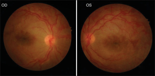
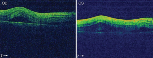
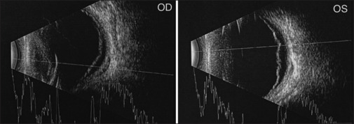
Purtscher retinopathy is a hemorrhagic and vaso-occlusive vasculopathy that results from head trauma. Typically, patients present with sudden onset of painless reduction in visual acuity and a group of retinal findings including retinal hemorrhages, retinal whitening, and optic disc edema. The objectives of the study were to describe the rare occurrence of combined central retinal artery and central vein occlusion in a child with Purtscher retinopathy and to illustrate the course of visual recovery and anatomical changes over 6 months of follow-up. The data were collected from the patient file including circumstances of presentation, visual acuity, anterior segment examination, fundus photography, fundus fluorescein angiography (FFA), B scan ultrasonography (B scan), and optical coherence tomography (OCT). Follow-ups over a 6-month period with repeated imaging were documented. A 6-year-old boy presented with a complaint of bilateral vision loss which occurred 2 days after falling out of high bed. On examination, visual acuity was hand motion bilaterally. External and anterior segments exhibited regular examination, apart from traumatic iritis in both eyes. Posterior segment examination showed intraretinal hemorrhages, retinal whitening bilaterally, and exudative retinal detachment, which was confirmed by OCT and B-scan. FFA of both the eyes showed delayed arterial and venous filling with macular hypoperfusion. Comprehensive systemic workup including brain imaging, hematology, immunology, and uveitis screening was negative. The patient was treated with a high-dose oral steroid. Six months later, his vision improved to 20/40 OD and 20/28 OS. Fundus examination and OCT showed peripheral retinal atrophy but preserved subfoveal outer retinal layers, which explained the visual improvement. Treatment with corticosteroids seems to be effective in reducing retinal edema and hastened visual recovery in patients with Purtscher retinopathy. Cilioretinal artery sparing can preserve the central vision in cases with combined artery and vein occlusion.
期刊介绍:
The Middle East African Journal of Ophthalmology (MEAJO), published four times per year in print and online, is an official journal of the Middle East African Council of Ophthalmology (MEACO). It is an international, peer-reviewed journal whose mission includes publication of original research of interest to ophthalmologists in the Middle East and Africa, and to provide readers with high quality educational review articles from world-renown experts. MEAJO, previously known as Middle East Journal of Ophthalmology (MEJO) was founded by Dr Akef El Maghraby in 1993.


 分享
分享
 求助内容:
求助内容: 应助结果提醒方式:
 扫码关注我们
