Mairead B Butler, Georgios Papageorgiou, Evangelos D Kanoulas, Vasiliki Voulgaridou, Hessel Wijkstra, Massimo Mischi, Christophe K Mannaerts, Steven McDougall, William Colin Duncan, Weiping Lu, Vassilis Sboros
{"title":"Mapping of prostate cancer microvascular patterns using super-resolution ultrasound imaging.","authors":"Mairead B Butler, Georgios Papageorgiou, Evangelos D Kanoulas, Vasiliki Voulgaridou, Hessel Wijkstra, Massimo Mischi, Christophe K Mannaerts, Steven McDougall, William Colin Duncan, Weiping Lu, Vassilis Sboros","doi":"10.1186/s41747-025-00561-6","DOIUrl":null,"url":null,"abstract":"<p><strong>Background: </strong>Super-resolution ultrasound imaging (SRUI) is a rapidly expanding field with the potential to impact cancer management. Image processing algorithms applied to contrast-enhanced ultrasound (CEUS) video data can track the path of the contrast agent and produce high-resolution maps of vascular networks. Our aim was to develop SRUI for mapping prostate vascular dynamics and to assess the feasibility of identifying vascular patterns associated with prostate cancer.</p><p><strong>Methods: </strong>Tracking algorithms for SRUI were developed using in silico data and validated in pre-clinical CEUS video collected from the sheep ovary. Algorithm performance was then assessed in a retrospective study of 54 image planes within 14 human prostates. CEUS data was collected for each plane, and regions of suspected cancer in each were identified from biopsy data.</p><p><strong>Results: </strong>Of three algorithms assessed, utilising vascular knowledge was found to be the most robust method. Regions of suspected cancer were associated with increased blood flow volume and speed while avascular regions were also identified. Ten scan planes had confirmed Gleason 7 cancer; of these 10 planes, 7 had distinct regions of fast and high-volume flow, while 6 had both avascular and high flow regions. The cancer-free planes had more consistent, low blood flow values across the plane.</p><p><strong>Conclusion: </strong>SRUI can be used to identify imaging biomarkers associated with vascular architecture and dynamics. These multiparameter biomarkers may be useful in pinpointing regions of significant prostate cancer.</p><p><strong>Relevance statement: </strong>Super-resolution ultrasound imaging can generate microvascular maps of the prostate, revealing tissue patterns and presenting significant potential for the identification of multiple biomarkers associated with the localisation of prostate cancer.</p><p><strong>Trial registration: </strong>Retrospectively registered NCT02831920, date 5/7/2016 https://www.</p><p><strong>Clinicaltrials: </strong>gov/study/NCT02831920 .</p><p><strong>Key points: </strong>An algorithm was developed and tested in synthetic pre-clinical and clinical data. Maps of blood vessels were created using contrast-enhanced ultrasound imaging. Specific presentations of vasculature at regions of prostate cancer have been identified.</p>","PeriodicalId":36926,"journal":{"name":"European Radiology Experimental","volume":"9 1","pages":"25"},"PeriodicalIF":3.6000,"publicationDate":"2025-02-20","publicationTypes":"Journal Article","fieldsOfStudy":null,"isOpenAccess":false,"openAccessPdf":"https://www.ncbi.nlm.nih.gov/pmc/articles/PMC11842657/pdf/","citationCount":"0","resultStr":null,"platform":"Semanticscholar","paperid":null,"PeriodicalName":"European Radiology Experimental","FirstCategoryId":"1085","ListUrlMain":"https://doi.org/10.1186/s41747-025-00561-6","RegionNum":0,"RegionCategory":null,"ArticlePicture":[],"TitleCN":null,"AbstractTextCN":null,"PMCID":null,"EPubDate":"","PubModel":"","JCR":"Q1","JCRName":"RADIOLOGY, NUCLEAR MEDICINE & MEDICAL IMAGING","Score":null,"Total":0}
引用次数: 0
Abstract
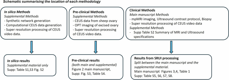
Background: Super-resolution ultrasound imaging (SRUI) is a rapidly expanding field with the potential to impact cancer management. Image processing algorithms applied to contrast-enhanced ultrasound (CEUS) video data can track the path of the contrast agent and produce high-resolution maps of vascular networks. Our aim was to develop SRUI for mapping prostate vascular dynamics and to assess the feasibility of identifying vascular patterns associated with prostate cancer.
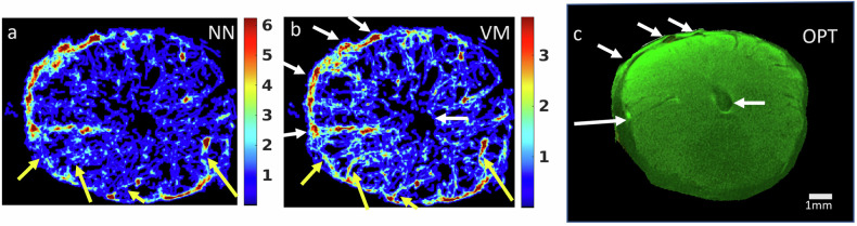
Methods: Tracking algorithms for SRUI were developed using in silico data and validated in pre-clinical CEUS video collected from the sheep ovary. Algorithm performance was then assessed in a retrospective study of 54 image planes within 14 human prostates. CEUS data was collected for each plane, and regions of suspected cancer in each were identified from biopsy data.
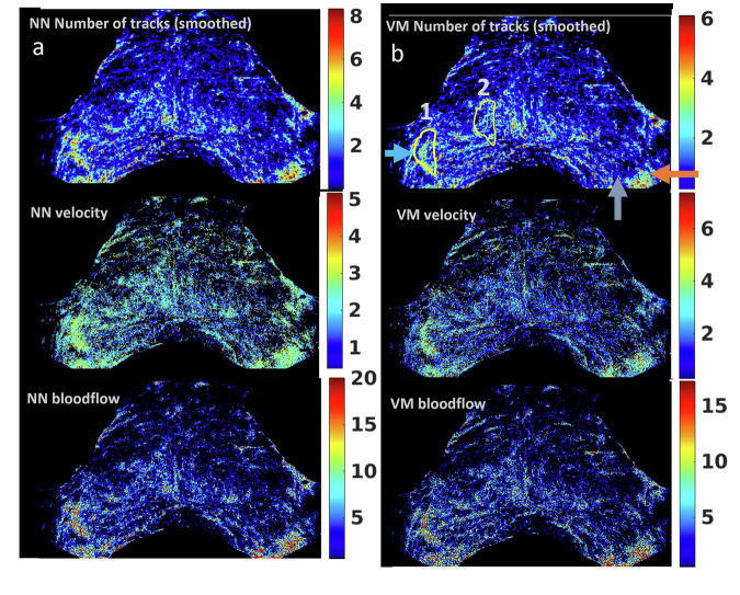
Results: Of three algorithms assessed, utilising vascular knowledge was found to be the most robust method. Regions of suspected cancer were associated with increased blood flow volume and speed while avascular regions were also identified. Ten scan planes had confirmed Gleason 7 cancer; of these 10 planes, 7 had distinct regions of fast and high-volume flow, while 6 had both avascular and high flow regions. The cancer-free planes had more consistent, low blood flow values across the plane.
Conclusion: SRUI can be used to identify imaging biomarkers associated with vascular architecture and dynamics. These multiparameter biomarkers may be useful in pinpointing regions of significant prostate cancer.
Relevance statement: Super-resolution ultrasound imaging can generate microvascular maps of the prostate, revealing tissue patterns and presenting significant potential for the identification of multiple biomarkers associated with the localisation of prostate cancer.
Trial registration: Retrospectively registered NCT02831920, date 5/7/2016 https://www.
Clinicaltrials: gov/study/NCT02831920 .
Key points: An algorithm was developed and tested in synthetic pre-clinical and clinical data. Maps of blood vessels were created using contrast-enhanced ultrasound imaging. Specific presentations of vasculature at regions of prostate cancer have been identified.


 分享
分享
 求助内容:
求助内容: 应助结果提醒方式:
 扫码关注我们
