Samra Pjanić, Goran Talić, Nikola Jevtić, Filip Golić, Ivan Soldatović, Nachiappan Chockalingam
{"title":"Ultrasound <i>vs.</i> x-ray: a new way for clinicians to track scoliosis progression?","authors":"Samra Pjanić, Goran Talić, Nikola Jevtić, Filip Golić, Ivan Soldatović, Nachiappan Chockalingam","doi":"10.4081/ejtm.2025.13422","DOIUrl":null,"url":null,"abstract":"<p><p>This retrospective study, utilising prospectively collected data, investigates the use of spine ultrasound as an alternative method for assessing scoliosis, with the aim of reducing radiation exposure. We included 92 patients aged 10 to 16 years with suspected idiopathic scoliosis. Exclusion criteria were weight over 150 kg, metal implants, pre-existing conditions, secondary deformities, and cognitive impairments. Each patient underwent clinical assessment and full spine radiographs, followed by spine ultrasound using the Scolioscan® system. Unprocessed B-mode ultrasound images were analysed using automatic measurements. The correlation between Ultrasound Coronal Angle (UCA) and Radiographic Cobb Angle (RCA) was evaluated at initial and follow-up visits. Strong correlations were found between UCA and RCA, with correlation coefficients ranging from 0.786 to 0.903 (p<0.001). The regression formula showed good predictive accuracy for curve progression on follow-up radiographs. The best results were observed in females and in primary thoracic curves (r = 0.936, p<0.001). Although only four patients exhibited true progression (≥5° increase in Cobb angle), changes in scoliotic angles were effectively detected using ultrasound. This study confirms the feasibility of unprocessed spine ultrasound for scoliosis monitoring in clinical settings. Automatic measurements without 3D reconstruction make ultrasound a practical tool for tracking progression. The regression model shows potential for predicting curve progression, although further validation is needed. These findings suggest spine ultrasound could reduce the need for radiographs, benefiting patients by minimising radiation exposure while providing reliable monitoring of scoliosis progression and treatment outcomes.</p>","PeriodicalId":46459,"journal":{"name":"European Journal of Translational Myology","volume":" ","pages":""},"PeriodicalIF":1.8000,"publicationDate":"2025-03-31","publicationTypes":"Journal Article","fieldsOfStudy":null,"isOpenAccess":false,"openAccessPdf":"https://www.ncbi.nlm.nih.gov/pmc/articles/PMC12038569/pdf/","citationCount":"0","resultStr":null,"platform":"Semanticscholar","paperid":null,"PeriodicalName":"European Journal of Translational Myology","FirstCategoryId":"1085","ListUrlMain":"https://doi.org/10.4081/ejtm.2025.13422","RegionNum":0,"RegionCategory":null,"ArticlePicture":[],"TitleCN":null,"AbstractTextCN":null,"PMCID":null,"EPubDate":"2025/2/21 0:00:00","PubModel":"Epub","JCR":"Q3","JCRName":"MEDICINE, RESEARCH & EXPERIMENTAL","Score":null,"Total":0}
引用次数: 0
Abstract
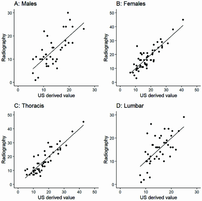
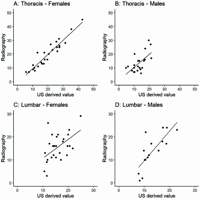
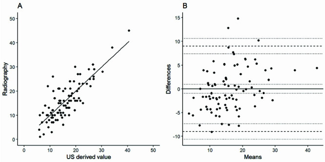
This retrospective study, utilising prospectively collected data, investigates the use of spine ultrasound as an alternative method for assessing scoliosis, with the aim of reducing radiation exposure. We included 92 patients aged 10 to 16 years with suspected idiopathic scoliosis. Exclusion criteria were weight over 150 kg, metal implants, pre-existing conditions, secondary deformities, and cognitive impairments. Each patient underwent clinical assessment and full spine radiographs, followed by spine ultrasound using the Scolioscan® system. Unprocessed B-mode ultrasound images were analysed using automatic measurements. The correlation between Ultrasound Coronal Angle (UCA) and Radiographic Cobb Angle (RCA) was evaluated at initial and follow-up visits. Strong correlations were found between UCA and RCA, with correlation coefficients ranging from 0.786 to 0.903 (p<0.001). The regression formula showed good predictive accuracy for curve progression on follow-up radiographs. The best results were observed in females and in primary thoracic curves (r = 0.936, p<0.001). Although only four patients exhibited true progression (≥5° increase in Cobb angle), changes in scoliotic angles were effectively detected using ultrasound. This study confirms the feasibility of unprocessed spine ultrasound for scoliosis monitoring in clinical settings. Automatic measurements without 3D reconstruction make ultrasound a practical tool for tracking progression. The regression model shows potential for predicting curve progression, although further validation is needed. These findings suggest spine ultrasound could reduce the need for radiographs, benefiting patients by minimising radiation exposure while providing reliable monitoring of scoliosis progression and treatment outcomes.


 分享
分享
 求助内容:
求助内容: 应助结果提醒方式:
 扫码关注我们
