Evaluation of the Influence of Changes in Bone Mineral Density and Increases in Articular Cartilage Thickness on Blood Supply of the Femoral Head in Legg-Calvé-Perthes Disease.
Hamid Reza Farpour, Mohammad Taghi Karimi, Mohammad Hossein Karimi
{"title":"Evaluation of the Influence of Changes in Bone Mineral Density and Increases in Articular Cartilage Thickness on Blood Supply of the Femoral Head in Legg-Calvé-Perthes Disease.","authors":"Hamid Reza Farpour, Mohammad Taghi Karimi, Mohammad Hossein Karimi","doi":"10.5371/hp.2025.37.1.38","DOIUrl":null,"url":null,"abstract":"<p><strong>Purpose: </strong>Although the etiology of Legg-Calvé-Perthes disease (LCPD) is not well understood, based on a new theory, it may be caused by a decrease in the supply of blood to the femoral head. The objective of this study was to examine the effects of a decrease in bone mineral density (BMD) and an increase in thickness of articular cartilage on the supply of blood to the femoral head in this group of patients.</p><p><strong>Materials and methods: </strong>This case study was based on a simulation analysis. Computed tomography scan images of a subject with Perthes disease were used to create a three-dimensional model of the hip joint on both the normal and Perthes sides. In addition, modeling of the blood vessels of the femoral head, including the foveolar and retinacular arteries, was performed during this study.</p><p><strong>Results: </strong>Increased stress on all articular components (femoral head, acetabulum, articular cartilage, and blood vessels) was observed on the Perthes side compared to the normal side. On the Perthes side with normal articular cartilage thickness, stress on all components, particularly the femur, showed a significant increase compared to the normal side.</p><p><strong>Conclusion: </strong>Increased deformation of the femoral head vessels was observed in patients with Perthes condition and when increased thickness of the articular cartilage was observed. A decrease in BMD can evidently increase the stress applied to the arteries of the femoral head, ultimately leading to death of the femoral head.</p>","PeriodicalId":73239,"journal":{"name":"Hip & pelvis","volume":"37 1","pages":"38-44"},"PeriodicalIF":0.0000,"publicationDate":"2025-03-01","publicationTypes":"Journal Article","fieldsOfStudy":null,"isOpenAccess":false,"openAccessPdf":"https://www.ncbi.nlm.nih.gov/pmc/articles/PMC11885784/pdf/","citationCount":"0","resultStr":null,"platform":"Semanticscholar","paperid":null,"PeriodicalName":"Hip & pelvis","FirstCategoryId":"1085","ListUrlMain":"https://doi.org/10.5371/hp.2025.37.1.38","RegionNum":0,"RegionCategory":null,"ArticlePicture":[],"TitleCN":null,"AbstractTextCN":null,"PMCID":null,"EPubDate":"","PubModel":"","JCR":"","JCRName":"","Score":null,"Total":0}
引用次数: 0
Abstract
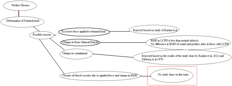
Purpose: Although the etiology of Legg-Calvé-Perthes disease (LCPD) is not well understood, based on a new theory, it may be caused by a decrease in the supply of blood to the femoral head. The objective of this study was to examine the effects of a decrease in bone mineral density (BMD) and an increase in thickness of articular cartilage on the supply of blood to the femoral head in this group of patients.
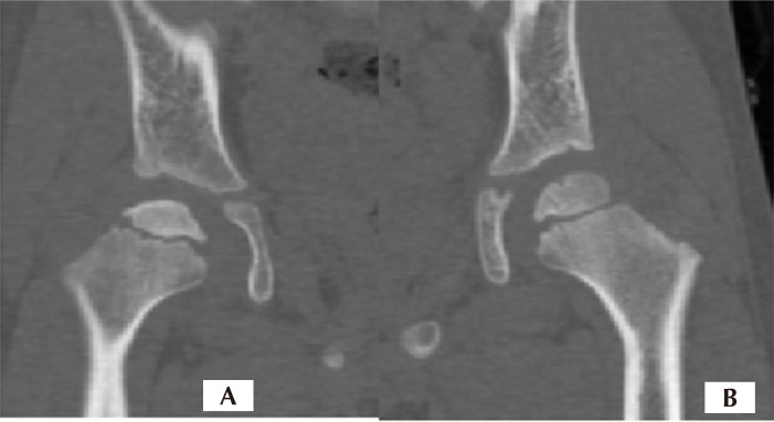
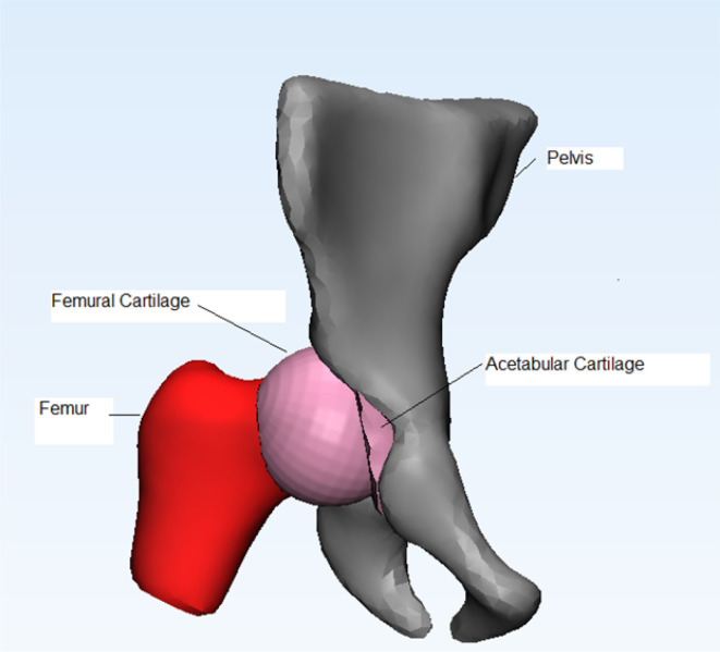
Materials and methods: This case study was based on a simulation analysis. Computed tomography scan images of a subject with Perthes disease were used to create a three-dimensional model of the hip joint on both the normal and Perthes sides. In addition, modeling of the blood vessels of the femoral head, including the foveolar and retinacular arteries, was performed during this study.
Results: Increased stress on all articular components (femoral head, acetabulum, articular cartilage, and blood vessels) was observed on the Perthes side compared to the normal side. On the Perthes side with normal articular cartilage thickness, stress on all components, particularly the femur, showed a significant increase compared to the normal side.
Conclusion: Increased deformation of the femoral head vessels was observed in patients with Perthes condition and when increased thickness of the articular cartilage was observed. A decrease in BMD can evidently increase the stress applied to the arteries of the femoral head, ultimately leading to death of the femoral head.


 分享
分享

 求助内容:
求助内容: 应助结果提醒方式:
 扫码关注我们
