Evaluation of the cortication ratio and visibility of mandibular canal and mandibular incisive canal in patients with mandibular cortical index type 1 on cone-beam computed tomography images.
Roghieh Bardal, Ahad Alizadeh, Vahid Nouri, Mohammad Salehi
{"title":"Evaluation of the cortication ratio and visibility of mandibular canal and mandibular incisive canal in patients with mandibular cortical index type 1 on cone-beam computed tomography images.","authors":"Roghieh Bardal, Ahad Alizadeh, Vahid Nouri, Mohammad Salehi","doi":"10.4103/jisp.jisp_275_23","DOIUrl":null,"url":null,"abstract":"<p><strong>Background: </strong>Mandibular canal visibility (MCV) is important to determine the relative position of the mandibular canal (MC) before any invasive surgery. It depends on the cortication ratio (CR) of the canal's superior border. This study aimed to evaluate the MCV and CR in patients with mandibular cortical index 1 (MCI1).</p><p><strong>Materials and methods: </strong>In this retrospective study, 132 mandibular cone-beam computed tomography images of patients with MCI1 were evaluated. 6-point rating MCV score and CR were determined for cross-sections of the MC in the following areas: incisive canal (INC), mental foramen and canal (MF), first premolar to the third molar (1PM, 2PM, 1M, 2M, and 3M), and the corresponding edentulous areas (E1PM-E3M). 1PM was overlapped with MF in most cases. An MCV score1 (excellent visibility) and 2PM area were considered reference levels.</p><p><strong>Results: </strong>Sex and age did not significantly affect the MCV score or CR (<i>P</i> > 0.05). 98.6% of the INC and 92.31%-100% of the MC were visible. The mean ± SD of the CR in the INC and MC was 0.86 (0.23) and 0.77 (0.29), respectively. The estimated difference in the mean CR was statistically significant only between the INC area and dentulous areas (<i>P</i> < 0.001).</p><p><strong>Conclusion: </strong>Despite the higher presence probability of score 2 in the 1M relative to 2PM, there was no significant decrease in the visible cases and CR of this area. Invisible cases were significantly lower in the INC, MF, and 3M areas.</p>","PeriodicalId":15890,"journal":{"name":"Journal of Indian Society of Periodontology","volume":"28 4","pages":"449-455"},"PeriodicalIF":0.0000,"publicationDate":"2024-07-01","publicationTypes":"Journal Article","fieldsOfStudy":null,"isOpenAccess":false,"openAccessPdf":"https://www.ncbi.nlm.nih.gov/pmc/articles/PMC11864344/pdf/","citationCount":"0","resultStr":null,"platform":"Semanticscholar","paperid":null,"PeriodicalName":"Journal of Indian Society of Periodontology","FirstCategoryId":"1085","ListUrlMain":"https://doi.org/10.4103/jisp.jisp_275_23","RegionNum":0,"RegionCategory":null,"ArticlePicture":[],"TitleCN":null,"AbstractTextCN":null,"PMCID":null,"EPubDate":"2025/1/6 0:00:00","PubModel":"Epub","JCR":"Q2","JCRName":"Dentistry","Score":null,"Total":0}
引用次数: 0
Abstract
Background: Mandibular canal visibility (MCV) is important to determine the relative position of the mandibular canal (MC) before any invasive surgery. It depends on the cortication ratio (CR) of the canal's superior border. This study aimed to evaluate the MCV and CR in patients with mandibular cortical index 1 (MCI1).
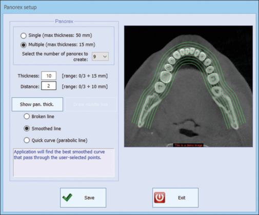
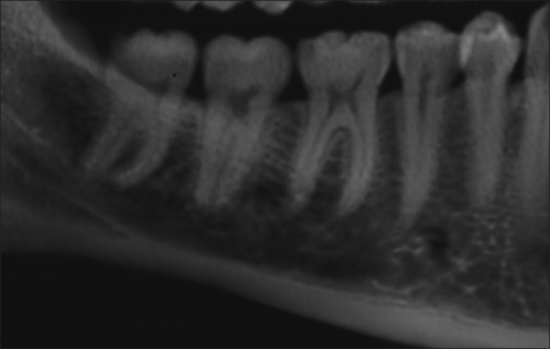
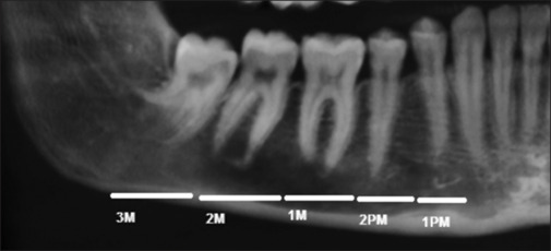
Materials and methods: In this retrospective study, 132 mandibular cone-beam computed tomography images of patients with MCI1 were evaluated. 6-point rating MCV score and CR were determined for cross-sections of the MC in the following areas: incisive canal (INC), mental foramen and canal (MF), first premolar to the third molar (1PM, 2PM, 1M, 2M, and 3M), and the corresponding edentulous areas (E1PM-E3M). 1PM was overlapped with MF in most cases. An MCV score1 (excellent visibility) and 2PM area were considered reference levels.
Results: Sex and age did not significantly affect the MCV score or CR (P > 0.05). 98.6% of the INC and 92.31%-100% of the MC were visible. The mean ± SD of the CR in the INC and MC was 0.86 (0.23) and 0.77 (0.29), respectively. The estimated difference in the mean CR was statistically significant only between the INC area and dentulous areas (P < 0.001).
Conclusion: Despite the higher presence probability of score 2 in the 1M relative to 2PM, there was no significant decrease in the visible cases and CR of this area. Invisible cases were significantly lower in the INC, MF, and 3M areas.
期刊介绍:
The Journal of Indian Society of Periodontology publishes original scientific articles to support practice , education and research in the dental specialty of periodontology and oral implantology. Journal of Indian Society of Periodontology (JISP), is the official publication of the Society and is managed and brought out by the Editor of the society. The journal is published Bimonthly with special issues being brought out for specific occasions. The ISP had a bulletin as its publication for a large number of years and was enhanced as a Journal a few years ago


 分享
分享
 求助内容:
求助内容: 应助结果提醒方式:
 扫码关注我们
