Yong Jun Jin, Jae-Young Park, Jun Young Chung, Sujin Noh, Hee-Woong Yun, Sumin Lim, Do Young Park
{"title":"McMurray's test is influenced by perimeniscal synovitis in degenerative meniscus tears.","authors":"Yong Jun Jin, Jae-Young Park, Jun Young Chung, Sujin Noh, Hee-Woong Yun, Sumin Lim, Do Young Park","doi":"10.1186/s43019-024-00242-5","DOIUrl":null,"url":null,"abstract":"<p><strong>Background: </strong>McMurray's test is a useful physical examination in determining meniscus tears, yet its sensitivity is only 38-62%. Furthermore, the relationship between degenerative meniscus tears (DMT) and mechanical symptoms during McMurray's test is not well defined. Perimeniscal synovitis occurs in osteoarthritic (OA) knees, inducing localized symptoms such as posterior knee pain in medial meniscus posterior horn DMTs. This study aimed to determine the relationship between McMurray's test with medial meniscus DMT and perimeniscal synovitis in patients with knee OA.</p><p><strong>Methods: </strong>We retrospectively analyzed 60 patients who underwent medial unicompartmental knee arthroplasty (UKA) with positive (n = 20) and negative (n = 40) preoperative McMurray's tests. Preoperative magnetic resonance imaging (MRI), intraoperative gross morphology, and histological analysis of meniscus and synovium were evaluated to determine meniscal tears and perimeniscal synovitis. Univariate and multivariate regression analyses were done to determine the effects of meniscus tears and synovitis on McMurray's test results.</p><p><strong>Results: </strong>Gross morphology of the medial meniscus (MM) showed 14 out of 20 torn menisci in the McMurray's (+) group compared with 22 out of 40 in the (-) group, with no difference in meniscus tear severity among groups. The (+) group showed higher values of synovial thickness (p < 0.001) and area (p < 0.001) compared with the (-) group on magnetic resonance imaging (MRI). Histological analysis showed higher synovitis (p < 0.001) scores and expression of inflammatory markers [interleukin (IL)-1β (p < 0.001), IL-6 (p = 0.007), nerve growth factor (NGF) (p = 0.003), inducible nitric oxide synthase (iNOS) (p < 0.001)] in the perimeniscal synovium of (+) group compared with the (-) group. Multivariable logistic analysis revealed that larger synovial area [odds ratio (OR) = 1.106, p = 0.008] and a higher histologic synovitis score (OR = 2.595, p = 0.011) were independently significant predictive factors for a positive McMurray's test.</p><p><strong>Conclusions: </strong>McMurray's test may be influenced by perimeniscal synovitis in DMT patients. The clinical implications of our results may influence not only the interpretation of McMurray's test but also the target tissue in treating mechanical symptoms related to meniscus tears.</p><p><strong>Level of evidence: </strong>Level II.</p>","PeriodicalId":36317,"journal":{"name":"Knee Surgery and Related Research","volume":"37 1","pages":"9"},"PeriodicalIF":4.4000,"publicationDate":"2025-02-28","publicationTypes":"Journal Article","fieldsOfStudy":null,"isOpenAccess":false,"openAccessPdf":"https://www.ncbi.nlm.nih.gov/pmc/articles/PMC11871708/pdf/","citationCount":"0","resultStr":null,"platform":"Semanticscholar","paperid":null,"PeriodicalName":"Knee Surgery and Related Research","FirstCategoryId":"1085","ListUrlMain":"https://doi.org/10.1186/s43019-024-00242-5","RegionNum":0,"RegionCategory":null,"ArticlePicture":[],"TitleCN":null,"AbstractTextCN":null,"PMCID":null,"EPubDate":"","PubModel":"","JCR":"Q2","JCRName":"Medicine","Score":null,"Total":0}
引用次数: 0
Abstract
Background: McMurray's test is a useful physical examination in determining meniscus tears, yet its sensitivity is only 38-62%. Furthermore, the relationship between degenerative meniscus tears (DMT) and mechanical symptoms during McMurray's test is not well defined. Perimeniscal synovitis occurs in osteoarthritic (OA) knees, inducing localized symptoms such as posterior knee pain in medial meniscus posterior horn DMTs. This study aimed to determine the relationship between McMurray's test with medial meniscus DMT and perimeniscal synovitis in patients with knee OA.
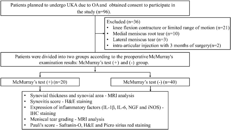
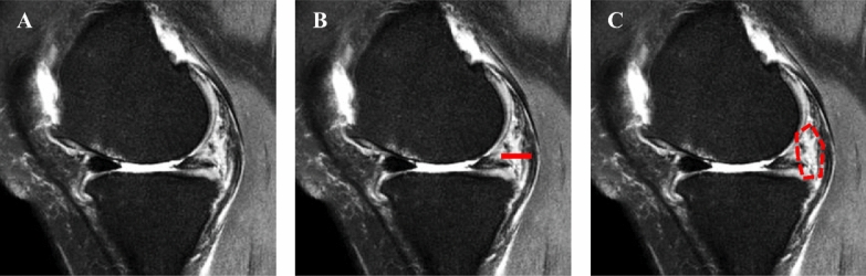
Methods: We retrospectively analyzed 60 patients who underwent medial unicompartmental knee arthroplasty (UKA) with positive (n = 20) and negative (n = 40) preoperative McMurray's tests. Preoperative magnetic resonance imaging (MRI), intraoperative gross morphology, and histological analysis of meniscus and synovium were evaluated to determine meniscal tears and perimeniscal synovitis. Univariate and multivariate regression analyses were done to determine the effects of meniscus tears and synovitis on McMurray's test results.
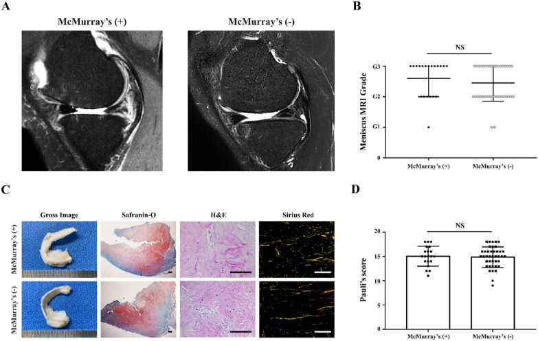
Results: Gross morphology of the medial meniscus (MM) showed 14 out of 20 torn menisci in the McMurray's (+) group compared with 22 out of 40 in the (-) group, with no difference in meniscus tear severity among groups. The (+) group showed higher values of synovial thickness (p < 0.001) and area (p < 0.001) compared with the (-) group on magnetic resonance imaging (MRI). Histological analysis showed higher synovitis (p < 0.001) scores and expression of inflammatory markers [interleukin (IL)-1β (p < 0.001), IL-6 (p = 0.007), nerve growth factor (NGF) (p = 0.003), inducible nitric oxide synthase (iNOS) (p < 0.001)] in the perimeniscal synovium of (+) group compared with the (-) group. Multivariable logistic analysis revealed that larger synovial area [odds ratio (OR) = 1.106, p = 0.008] and a higher histologic synovitis score (OR = 2.595, p = 0.011) were independently significant predictive factors for a positive McMurray's test.
Conclusions: McMurray's test may be influenced by perimeniscal synovitis in DMT patients. The clinical implications of our results may influence not only the interpretation of McMurray's test but also the target tissue in treating mechanical symptoms related to meniscus tears.


 分享
分享
 求助内容:
求助内容: 应助结果提醒方式:
 扫码关注我们
