{"title":"Transient Absorption Microscopy for Early-Stage Imaging of Protein Aggregates Using Thioflavin T","authors":"Przemyslaw Slota, Piotr Fita","doi":"10.1021/acsphotonics.4c01900","DOIUrl":null,"url":null,"abstract":"We present the design and application of a transient absorption microscope for imaging protein aggregates at early stages of growth, utilizing the widely used amyloid marker dye Thioflavin T (ThT). By employing femtosecond laser pulses for excitation and probing, the microscope distinguishes signals from free ThT molecules and ThT bound to protein aggregates, enabling the imaging of aggregates at different stages of development. Spatially resolved transient absorption measurements revealed two distinct excited-state lifetimes for ThT in the presence of insulin aggregates: a short lifetime (approximately 10 ps) and a longer lifetime (in the range of 100–200 ps). The short lifetime dominates at early stages of aggregation, reflecting ThT binding to small, disordered aggregates or amorphous precursors. As aggregation progresses, the contribution of the longer lifetime increases, corresponding to the formation of larger, more ordered structures. This observation agrees with theoretical models of amyloid formation, where early aggregates serve as nucleation sites for the growth of mature fibrils. The results demonstrate the capability of transient absorption microscopy to resolve spatial and temporal dynamics of protein aggregation. This approach offers potential applications in the study of amyloid-related diseases, including the characterization of aggregation inhibitors and imaging of pathological protein deposits in tissue samples.","PeriodicalId":23,"journal":{"name":"ACS Photonics","volume":"86 1","pages":""},"PeriodicalIF":6.7000,"publicationDate":"2025-03-05","publicationTypes":"Journal Article","fieldsOfStudy":null,"isOpenAccess":false,"openAccessPdf":"","citationCount":"0","resultStr":null,"platform":"Semanticscholar","paperid":null,"PeriodicalName":"ACS Photonics","FirstCategoryId":"101","ListUrlMain":"https://doi.org/10.1021/acsphotonics.4c01900","RegionNum":1,"RegionCategory":"物理与天体物理","ArticlePicture":[],"TitleCN":null,"AbstractTextCN":null,"PMCID":null,"EPubDate":"","PubModel":"","JCR":"Q1","JCRName":"MATERIALS SCIENCE, MULTIDISCIPLINARY","Score":null,"Total":0}
引用次数: 0
Abstract
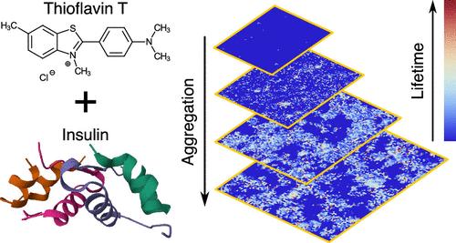
We present the design and application of a transient absorption microscope for imaging protein aggregates at early stages of growth, utilizing the widely used amyloid marker dye Thioflavin T (ThT). By employing femtosecond laser pulses for excitation and probing, the microscope distinguishes signals from free ThT molecules and ThT bound to protein aggregates, enabling the imaging of aggregates at different stages of development. Spatially resolved transient absorption measurements revealed two distinct excited-state lifetimes for ThT in the presence of insulin aggregates: a short lifetime (approximately 10 ps) and a longer lifetime (in the range of 100–200 ps). The short lifetime dominates at early stages of aggregation, reflecting ThT binding to small, disordered aggregates or amorphous precursors. As aggregation progresses, the contribution of the longer lifetime increases, corresponding to the formation of larger, more ordered structures. This observation agrees with theoretical models of amyloid formation, where early aggregates serve as nucleation sites for the growth of mature fibrils. The results demonstrate the capability of transient absorption microscopy to resolve spatial and temporal dynamics of protein aggregation. This approach offers potential applications in the study of amyloid-related diseases, including the characterization of aggregation inhibitors and imaging of pathological protein deposits in tissue samples.
期刊介绍:
Published as soon as accepted and summarized in monthly issues, ACS Photonics will publish Research Articles, Letters, Perspectives, and Reviews, to encompass the full scope of published research in this field.


 分享
分享
 求助内容:
求助内容: 应助结果提醒方式:
 扫码关注我们
