V M Vimala Priyadharshini, Indirani Muthukrishnan, Dinesh Kumar Gauthaman, Shelley Simon
{"title":"[18F]FDG- PET/CT in Diagnosis, Staging, and Management of Patients with Langerhans Cell Histiocytosis.","authors":"V M Vimala Priyadharshini, Indirani Muthukrishnan, Dinesh Kumar Gauthaman, Shelley Simon","doi":"10.4103/ijnm.ijnm_46_24","DOIUrl":null,"url":null,"abstract":"<p><strong>Background: </strong>Langerhans cell histiocytosis (LCH), a rare hematological disorder, presents significant diagnostic challenges due to its varied clinical manifestations. This study aims to analyse the use of F-18 fluoro-deoxy-glucose positron emission tomography computed tomography (F-18 FDG PET/CT) in diagnosis, staging, and management of LCH.</p><p><strong>Materials and methods: </strong>Fifty-nine patients with LCH were included, who underwent a total of ninety-three F-18 FDG PET/CT scans (including follow-up scans in 19 patients). The sites of abnormal FDG uptake were assessed and the maximum standardized uptake value was measured in all the scans.</p><p><strong>Results: </strong>Twenty-five patients (42.4%) had single system LCH (SS-LCH) and 34 patients (57.6%) had multisystem involvement LCH, 49/59. The most common sites of LCH involvement were bones (49/59, 83.1%) and lymph nodes (39/59, 44.9%). 12/59 patients (20.3%) had unifocal SS-LCH bone lesions, mostly in skull. The other common sites involved were lungs, liver, spleen, marrow, skin, and soft tissues. Less commonly involved sites included pancreas (2 cases), occipital lobe (1 case), and bowel (1 case). PET/CT was used in response assessment in 19 patients and helped in initiation of second line chemotherapy in cases of disease progression (2 cases) and relapse (2 cases). Seven cases with clinical suspicion were diagnosed as LCH based on lesion characteristics and FDG uptake, which were later biopsy proven.</p><p><strong>Conclusion: </strong>F-18 FDG PET/CT revealed morphological and metabolic characteristics of LCH lesions, aiding in accurate diagnosis, assessment of disease burden, and prognostication, thereby can be used as a comprehensive imaging tool in management of LCH.</p>","PeriodicalId":45830,"journal":{"name":"Indian Journal of Nuclear Medicine","volume":"39 5","pages":"341-346"},"PeriodicalIF":0.5000,"publicationDate":"2024-09-01","publicationTypes":"Journal Article","fieldsOfStudy":null,"isOpenAccess":false,"openAccessPdf":"https://www.ncbi.nlm.nih.gov/pmc/articles/PMC11884357/pdf/","citationCount":"0","resultStr":null,"platform":"Semanticscholar","paperid":null,"PeriodicalName":"Indian Journal of Nuclear Medicine","FirstCategoryId":"1085","ListUrlMain":"https://doi.org/10.4103/ijnm.ijnm_46_24","RegionNum":0,"RegionCategory":null,"ArticlePicture":[],"TitleCN":null,"AbstractTextCN":null,"PMCID":null,"EPubDate":"2025/1/25 0:00:00","PubModel":"Epub","JCR":"Q4","JCRName":"RADIOLOGY, NUCLEAR MEDICINE & MEDICAL IMAGING","Score":null,"Total":0}
引用次数: 0
Abstract
Background: Langerhans cell histiocytosis (LCH), a rare hematological disorder, presents significant diagnostic challenges due to its varied clinical manifestations. This study aims to analyse the use of F-18 fluoro-deoxy-glucose positron emission tomography computed tomography (F-18 FDG PET/CT) in diagnosis, staging, and management of LCH.
Materials and methods: Fifty-nine patients with LCH were included, who underwent a total of ninety-three F-18 FDG PET/CT scans (including follow-up scans in 19 patients). The sites of abnormal FDG uptake were assessed and the maximum standardized uptake value was measured in all the scans.
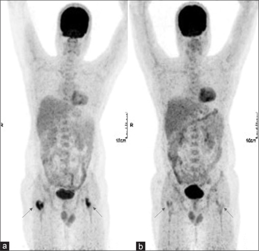
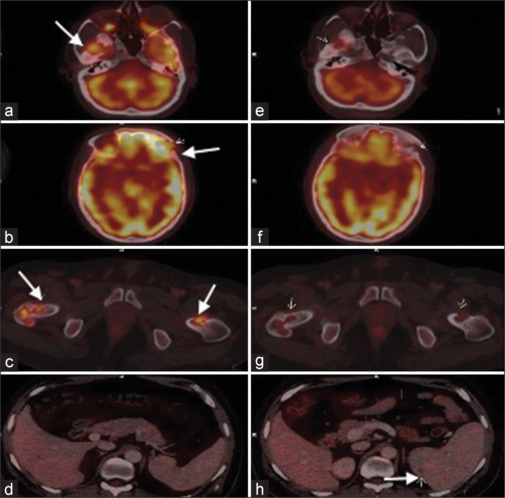
Results: Twenty-five patients (42.4%) had single system LCH (SS-LCH) and 34 patients (57.6%) had multisystem involvement LCH, 49/59. The most common sites of LCH involvement were bones (49/59, 83.1%) and lymph nodes (39/59, 44.9%). 12/59 patients (20.3%) had unifocal SS-LCH bone lesions, mostly in skull. The other common sites involved were lungs, liver, spleen, marrow, skin, and soft tissues. Less commonly involved sites included pancreas (2 cases), occipital lobe (1 case), and bowel (1 case). PET/CT was used in response assessment in 19 patients and helped in initiation of second line chemotherapy in cases of disease progression (2 cases) and relapse (2 cases). Seven cases with clinical suspicion were diagnosed as LCH based on lesion characteristics and FDG uptake, which were later biopsy proven.
Conclusion: F-18 FDG PET/CT revealed morphological and metabolic characteristics of LCH lesions, aiding in accurate diagnosis, assessment of disease burden, and prognostication, thereby can be used as a comprehensive imaging tool in management of LCH.


 分享
分享
 求助内容:
求助内容: 应助结果提醒方式:
 扫码关注我们
