{"title":"FDG PET Scan in Cutaneous Rosai-Dorfman-Destombes Disease.","authors":"Shrikant Vasantrao Solav, Chakor Vora, Rajlaxmi Jagtap, Shailendra Savale, Ishtiyaq Shaikh, Aman Solav","doi":"10.4103/ijnm.ijnm_121_24","DOIUrl":null,"url":null,"abstract":"<p><p>Rosai-Dorfman-Destombes (RDD) disease is also called as sinus histiocytosis and is characterized by enlarged lymph nodes and previously called as non-Langerhans cell histiocytosis. Based on pathologic, molecular, and genetic features, RDD disease has been classified into sporadic noncutaneous (classical nodal, extranodal, neoplasia associated, and autoimmune associated), familial (H syndrome, autoimmune lymphoproliferative syndrome related, and familial NOS), and cutaneous subtypes. Cutaneous RDD disease is not associated with lymphadenopathy or visceral organ involvement. The disease is usually localized and has relatively better long-term prognosis. Presented here is a case of indurated plaque-like skin lesions over the abdomen. <sup>18</sup>F-fluorodeoxyglucose (FDG) positron emission tomography-computed tomography scan revealed FDG avid cutaneous-subcutaneous soft-tissue lesions. Histology confirmed the diagnosis of cutaneous RDD disease.</p>","PeriodicalId":45830,"journal":{"name":"Indian Journal of Nuclear Medicine","volume":"39 5","pages":"396-398"},"PeriodicalIF":0.5000,"publicationDate":"2024-09-01","publicationTypes":"Journal Article","fieldsOfStudy":null,"isOpenAccess":false,"openAccessPdf":"https://www.ncbi.nlm.nih.gov/pmc/articles/PMC11884345/pdf/","citationCount":"0","resultStr":null,"platform":"Semanticscholar","paperid":null,"PeriodicalName":"Indian Journal of Nuclear Medicine","FirstCategoryId":"1085","ListUrlMain":"https://doi.org/10.4103/ijnm.ijnm_121_24","RegionNum":0,"RegionCategory":null,"ArticlePicture":[],"TitleCN":null,"AbstractTextCN":null,"PMCID":null,"EPubDate":"2025/1/25 0:00:00","PubModel":"Epub","JCR":"Q4","JCRName":"RADIOLOGY, NUCLEAR MEDICINE & MEDICAL IMAGING","Score":null,"Total":0}
引用次数: 0
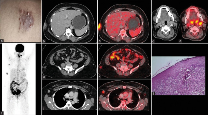
Abstract
Rosai-Dorfman-Destombes (RDD) disease is also called as sinus histiocytosis and is characterized by enlarged lymph nodes and previously called as non-Langerhans cell histiocytosis. Based on pathologic, molecular, and genetic features, RDD disease has been classified into sporadic noncutaneous (classical nodal, extranodal, neoplasia associated, and autoimmune associated), familial (H syndrome, autoimmune lymphoproliferative syndrome related, and familial NOS), and cutaneous subtypes. Cutaneous RDD disease is not associated with lymphadenopathy or visceral organ involvement. The disease is usually localized and has relatively better long-term prognosis. Presented here is a case of indurated plaque-like skin lesions over the abdomen. 18F-fluorodeoxyglucose (FDG) positron emission tomography-computed tomography scan revealed FDG avid cutaneous-subcutaneous soft-tissue lesions. Histology confirmed the diagnosis of cutaneous RDD disease.


 分享
分享
 求助内容:
求助内容: 应助结果提醒方式:
 扫码关注我们
