Tugcan Alp Kirkizlar, Onur Kirkizlar, Selin Soyluoglu, Elif Gulsum Umit, Funda Ustun, Ahmet Muzaffer Demir
{"title":"The Impact of Focal Lesions on Overall and Progression-free Survival in Multiple Myeloma.","authors":"Tugcan Alp Kirkizlar, Onur Kirkizlar, Selin Soyluoglu, Elif Gulsum Umit, Funda Ustun, Ahmet Muzaffer Demir","doi":"10.4103/ijnm.ijnm_131_24","DOIUrl":null,"url":null,"abstract":"<p><strong>Purpose: </strong>In this study, we aimed to reveal the incidence of ≥3 focal lesions (FLs) and analyze overall survival (OS) and progression-free survival (PFS) according to the number of FLs, as well as to identify mortality and PFS risk factors, in our newly diagnosed multiple myeloma (NDMM) patients.</p><p><strong>Materials and methods: </strong>A total of 89 NDMM patients who underwent <sup>18</sup>F-FDG positron emission tomography/computerized tomography (PET/CT) imaging were included in the study.</p><p><strong>Results: </strong>While 57.3% of the patients had ≥3 FLs, 20.2% had no FL. The median OS and PFS were 55 and 43 months, respectively. The median survival time was 49 months for patients with ≥3 FLs and 101 months for patients with <3 FLs, with a statistically significant difference (<i>P</i> = 0.049). The median PFS was 34 months in patients with ≥3 FLs and 67 months in patients with <3 FLs; this difference was also statistically significant (<i>P</i> = 0.026). The difference in median survival was statistically significant, based on whether autologous stem cell transplantation (ASCT) was performed and the number of FLs (≥3 or <3) (<i>P</i> = 0.011). In the multivariate regression analysis, ≥3 FLs was not a predictor of mortality but was a risk factor for PFS.</p><p><strong>Conclusion: </strong>In our study, we observed significantly worse OS and PFS in patients with ≥3 FLs at diagnosis, and it is noteworthy that the OS was worse in those patients who did not undergo ASCT. <sup>18</sup>F-FDG PET/CT is a feasible imaging technique for the prediction of prognosis in the initial evaluation of NDMM, and we believe that consolidation with ASCT as a modifiable factor, especially in patients with ≥3 FLs, will lead to a more favorable prognosis.</p>","PeriodicalId":45830,"journal":{"name":"Indian Journal of Nuclear Medicine","volume":"39 5","pages":"353-359"},"PeriodicalIF":0.5000,"publicationDate":"2024-09-01","publicationTypes":"Journal Article","fieldsOfStudy":null,"isOpenAccess":false,"openAccessPdf":"https://www.ncbi.nlm.nih.gov/pmc/articles/PMC11884343/pdf/","citationCount":"0","resultStr":null,"platform":"Semanticscholar","paperid":null,"PeriodicalName":"Indian Journal of Nuclear Medicine","FirstCategoryId":"1085","ListUrlMain":"https://doi.org/10.4103/ijnm.ijnm_131_24","RegionNum":0,"RegionCategory":null,"ArticlePicture":[],"TitleCN":null,"AbstractTextCN":null,"PMCID":null,"EPubDate":"2025/1/25 0:00:00","PubModel":"Epub","JCR":"Q4","JCRName":"RADIOLOGY, NUCLEAR MEDICINE & MEDICAL IMAGING","Score":null,"Total":0}
引用次数: 0
Abstract
Purpose: In this study, we aimed to reveal the incidence of ≥3 focal lesions (FLs) and analyze overall survival (OS) and progression-free survival (PFS) according to the number of FLs, as well as to identify mortality and PFS risk factors, in our newly diagnosed multiple myeloma (NDMM) patients.
Materials and methods: A total of 89 NDMM patients who underwent 18F-FDG positron emission tomography/computerized tomography (PET/CT) imaging were included in the study.
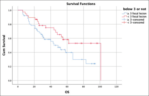
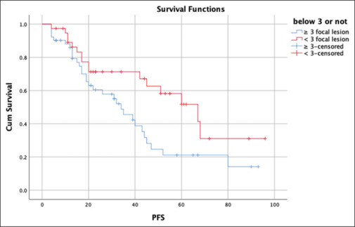
Results: While 57.3% of the patients had ≥3 FLs, 20.2% had no FL. The median OS and PFS were 55 and 43 months, respectively. The median survival time was 49 months for patients with ≥3 FLs and 101 months for patients with <3 FLs, with a statistically significant difference (P = 0.049). The median PFS was 34 months in patients with ≥3 FLs and 67 months in patients with <3 FLs; this difference was also statistically significant (P = 0.026). The difference in median survival was statistically significant, based on whether autologous stem cell transplantation (ASCT) was performed and the number of FLs (≥3 or <3) (P = 0.011). In the multivariate regression analysis, ≥3 FLs was not a predictor of mortality but was a risk factor for PFS.
Conclusion: In our study, we observed significantly worse OS and PFS in patients with ≥3 FLs at diagnosis, and it is noteworthy that the OS was worse in those patients who did not undergo ASCT. 18F-FDG PET/CT is a feasible imaging technique for the prediction of prognosis in the initial evaluation of NDMM, and we believe that consolidation with ASCT as a modifiable factor, especially in patients with ≥3 FLs, will lead to a more favorable prognosis.


 分享
分享
 求助内容:
求助内容: 应助结果提醒方式:
 扫码关注我们
