Nuan Lin, Koen C van Zomeren, Torsten Plosch, Naomi Hofsink, Teelkien van Veen, Hui Ting Li, Jiazhe Lin, Xiaoling Zhou, Henk Groen, Uwe J F Tietge, Astrid Cantineau, Romana Schirhagl, Annemieke Hoek
{"title":"Follicle-stimulating hormone stimulates free radical generation without inducing substantial oxidative stress in human granulosa cells.","authors":"Nuan Lin, Koen C van Zomeren, Torsten Plosch, Naomi Hofsink, Teelkien van Veen, Hui Ting Li, Jiazhe Lin, Xiaoling Zhou, Henk Groen, Uwe J F Tietge, Astrid Cantineau, Romana Schirhagl, Annemieke Hoek","doi":"10.1093/hropen/hoaf007","DOIUrl":null,"url":null,"abstract":"<p><strong>Study question: </strong>Does FSH induce free radical generation with substantial oxidative damage in human cumulus granulosa cells (cGCs) and mural granulosa cells (mGCs)?</p><p><strong>Summary answer: </strong>FSH of both physiological and supraphysiological concentrations induced free radical generation on subcellular levels, most notably in the mitochondria, while the elevated free radical load caused neglectable oxidative damage in both cGCs and mGCs.</p><p><strong>What is known already: </strong>FSH is fundamental for regulation of granulosa cell (GC) function and oocyte maturation, during which a physiological level of reactive oxygen species (ROS) is essential, while excessive amounts lead to oxidative damage. Potential adverse effects of high FSH doses on GCs may be mediated by ROS.</p><p><strong>Study design size duration: </strong>This prospective experimental study included patients who attended a reproductive medicine center in 2023. cGC and mGC were separately isolated and brought into culture on the day of oocyte retrieval, 36 h after ovulation induction with recombinant hCG (250 mg). Recombinant FSH, at different concentrations, mimicking physiological (6 mIU/ml) and supraphysiological (60 and 600 mIU/ml) conditions, was applied (n = 4 in each group).</p><p><strong>Participants/materials setting methods: </strong>Women aged 20-35 years, undergoing ICSI with at least three follicles, were included. Quantum sensing of cGC and mGC free radicals, detected by either cytoplasm-located fluorescent nanodiamonds (FNDs) or mitochondria-targeted FNDs, was tracked for 2 h following FSH treatment in a magnetometry setup. Mitochondrial function analysis, as well as oxidative damage to DNA/RNA, lipids, and proteins, upon FSH exposure, was examined.</p><p><strong>Main results and the role of chance: </strong>FSH-induced cytoplasmic and mitochondrial ROS increases in cGC and mGC (<i>P</i> < 0.01 in all concentrations after 2 h) while showing different patterns along time: cGC showed significantly larger cytoplasmic ROS change compared with mGC to physiological (<i>P</i> < 0.01) and supraphysiological (<i>P</i> < 0.05) concentrations of FSH. Significantly larger free radical changes were observed in the mitochondria compared to the cytoplasm in response to FSH (all concentrations in cGCs with <i>P</i> < 0.05; supraphysiological concentrations in mGCs with <i>P</i> < 0.05, <i>P</i> < 0.001, respectively) after 2 h. Mitochondrial basal respiration and ATP production were significantly increased upon FSH exposure to supraphysiological concentrations in both cGCs (<i>P</i> < 0.01) and mGCs (<i>P</i> < 0.05). However, no oxidative damage to GC DNA/RNA, proteins, or lipids was found upon FSH exposure at any concentration except elevated lipid peroxidation in the FSH group of 600 mIU/ml (<i>P</i> < 0.05).</p><p><strong>Large scale data: </strong>N/A.</p><p><strong>Limitations reasons for caution: </strong>The GCs came from females of different biological backgrounds and were stimulated before oocyte and GC retrieval, thereby increasing the risk of variation. In addition, the effects of long-term FSH exposure as well as the effects of the FSH-induced ROS on the oocyte remain to be investigated.</p><p><strong>Wider implications of the findings: </strong>We demonstrate that FSH of both physiological and supraphysiological concentrations induces free radical generation at subcellular levels, most notably in the mitochondria, while the elevated free radical load causes neglectable oxidative damage in both cGCs and mGCs. Our results suggest that the 'FSH Ootoxicity' hypothesis would not seem to be mediated by ROS in human GCs.</p><p><strong>Study funding/competing interests: </strong>This study is supported by the Abel Tasman Talent Program (ATTP) of the Graduate School of Medical Sciences of the University Medical Center Groningen/University of Groningen, The Netherlands, as well as an XS grant from NWO. Unrelated to the current work, A.H. is a member of an advisory board on the development and application of a lifestyle App for patients with infertility from Ferring Pharmaceutical Company, The Netherlands. R.S. is the founder of QT Sense B.V., who commercialize quantum sensing equipment. This article has no direct relation to the work of QT Sense B.V. The remaining authors have no conflicts of interest.</p>","PeriodicalId":73264,"journal":{"name":"Human reproduction open","volume":"2025 2","pages":"hoaf007"},"PeriodicalIF":11.1000,"publicationDate":"2025-02-17","publicationTypes":"Journal Article","fieldsOfStudy":null,"isOpenAccess":false,"openAccessPdf":"https://www.ncbi.nlm.nih.gov/pmc/articles/PMC11893154/pdf/","citationCount":"0","resultStr":null,"platform":"Semanticscholar","paperid":null,"PeriodicalName":"Human reproduction open","FirstCategoryId":"1085","ListUrlMain":"https://doi.org/10.1093/hropen/hoaf007","RegionNum":0,"RegionCategory":null,"ArticlePicture":[],"TitleCN":null,"AbstractTextCN":null,"PMCID":null,"EPubDate":"2025/1/1 0:00:00","PubModel":"eCollection","JCR":"Q1","JCRName":"OBSTETRICS & GYNECOLOGY","Score":null,"Total":0}
引用次数: 0
Abstract
Study question: Does FSH induce free radical generation with substantial oxidative damage in human cumulus granulosa cells (cGCs) and mural granulosa cells (mGCs)?
Summary answer: FSH of both physiological and supraphysiological concentrations induced free radical generation on subcellular levels, most notably in the mitochondria, while the elevated free radical load caused neglectable oxidative damage in both cGCs and mGCs.
What is known already: FSH is fundamental for regulation of granulosa cell (GC) function and oocyte maturation, during which a physiological level of reactive oxygen species (ROS) is essential, while excessive amounts lead to oxidative damage. Potential adverse effects of high FSH doses on GCs may be mediated by ROS.
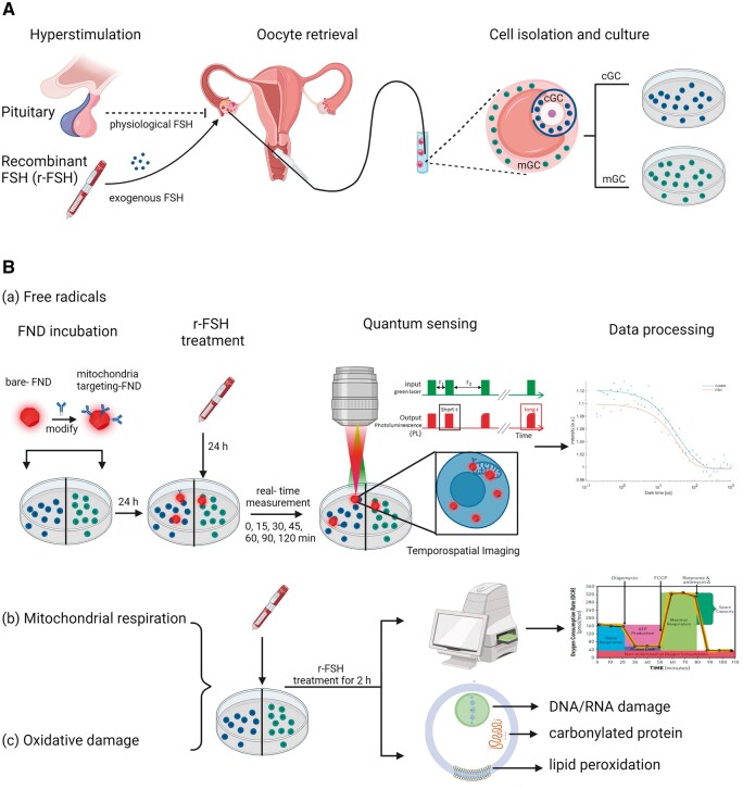
Study design size duration: This prospective experimental study included patients who attended a reproductive medicine center in 2023. cGC and mGC were separately isolated and brought into culture on the day of oocyte retrieval, 36 h after ovulation induction with recombinant hCG (250 mg). Recombinant FSH, at different concentrations, mimicking physiological (6 mIU/ml) and supraphysiological (60 and 600 mIU/ml) conditions, was applied (n = 4 in each group).
Participants/materials setting methods: Women aged 20-35 years, undergoing ICSI with at least three follicles, were included. Quantum sensing of cGC and mGC free radicals, detected by either cytoplasm-located fluorescent nanodiamonds (FNDs) or mitochondria-targeted FNDs, was tracked for 2 h following FSH treatment in a magnetometry setup. Mitochondrial function analysis, as well as oxidative damage to DNA/RNA, lipids, and proteins, upon FSH exposure, was examined.
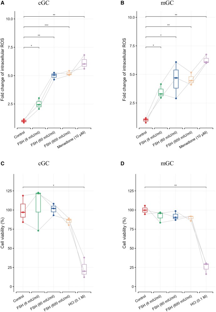
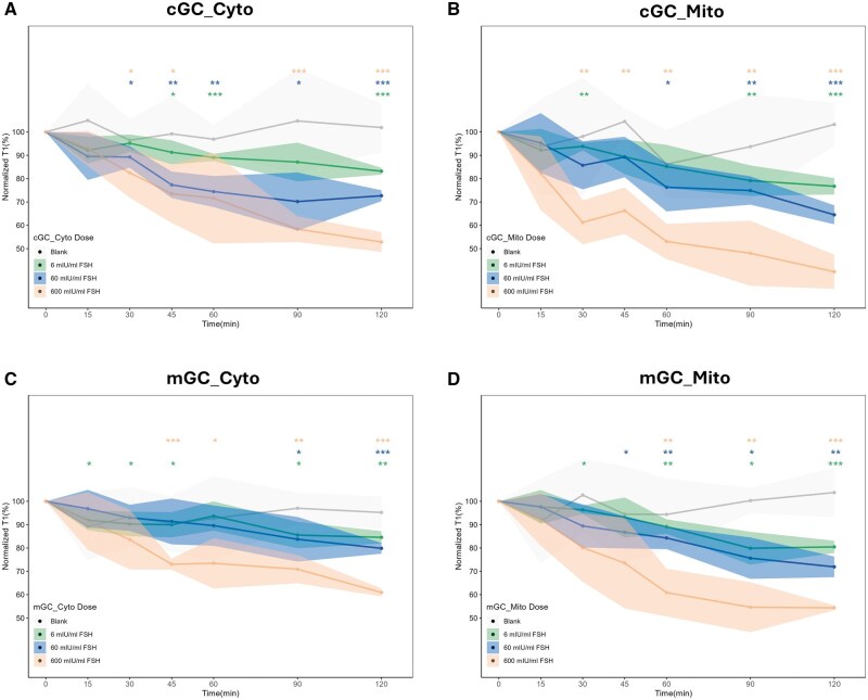
Main results and the role of chance: FSH-induced cytoplasmic and mitochondrial ROS increases in cGC and mGC (P < 0.01 in all concentrations after 2 h) while showing different patterns along time: cGC showed significantly larger cytoplasmic ROS change compared with mGC to physiological (P < 0.01) and supraphysiological (P < 0.05) concentrations of FSH. Significantly larger free radical changes were observed in the mitochondria compared to the cytoplasm in response to FSH (all concentrations in cGCs with P < 0.05; supraphysiological concentrations in mGCs with P < 0.05, P < 0.001, respectively) after 2 h. Mitochondrial basal respiration and ATP production were significantly increased upon FSH exposure to supraphysiological concentrations in both cGCs (P < 0.01) and mGCs (P < 0.05). However, no oxidative damage to GC DNA/RNA, proteins, or lipids was found upon FSH exposure at any concentration except elevated lipid peroxidation in the FSH group of 600 mIU/ml (P < 0.05).
Large scale data: N/A.
Limitations reasons for caution: The GCs came from females of different biological backgrounds and were stimulated before oocyte and GC retrieval, thereby increasing the risk of variation. In addition, the effects of long-term FSH exposure as well as the effects of the FSH-induced ROS on the oocyte remain to be investigated.
Wider implications of the findings: We demonstrate that FSH of both physiological and supraphysiological concentrations induces free radical generation at subcellular levels, most notably in the mitochondria, while the elevated free radical load causes neglectable oxidative damage in both cGCs and mGCs. Our results suggest that the 'FSH Ootoxicity' hypothesis would not seem to be mediated by ROS in human GCs.
Study funding/competing interests: This study is supported by the Abel Tasman Talent Program (ATTP) of the Graduate School of Medical Sciences of the University Medical Center Groningen/University of Groningen, The Netherlands, as well as an XS grant from NWO. Unrelated to the current work, A.H. is a member of an advisory board on the development and application of a lifestyle App for patients with infertility from Ferring Pharmaceutical Company, The Netherlands. R.S. is the founder of QT Sense B.V., who commercialize quantum sensing equipment. This article has no direct relation to the work of QT Sense B.V. The remaining authors have no conflicts of interest.
研究问题:FSH是否会诱导自由基生成,并对人积云颗粒细胞(cgc)和壁颗粒细胞(mGCs)造成实质性的氧化损伤?总结性回答:生理和超生理浓度的FSH诱导亚细胞水平的自由基生成,最明显的是在线粒体中,而升高的自由基负荷在cgc和mGCs中引起可忽略的氧化损伤。已知情况:卵泡刺激素是调节颗粒细胞(GC)功能和卵母细胞成熟的基础,在此过程中,生理水平的活性氧(ROS)是必不可少的,而过量会导致氧化损伤。高剂量FSH对GCs的潜在不良影响可能是由ROS介导的。研究设计规模持续时间:这项前瞻性实验研究包括2023年在生殖医学中心就诊的患者。重组hCG (250 mg)诱导排卵后36 h,取卵当天分别分离cGC和mGC进行培养。采用不同浓度的重组FSH,模拟生理(6 mIU/ml)和超生理(60和600 mIU/ml)条件(每组n = 4)。参与者/材料设置方法:年龄20-35岁,接受ICSI的女性,至少有三个卵泡。通过细胞质定位荧光纳米金刚石(FNDs)或线粒体靶向纳米金刚石(FNDs)检测cGC和mGC自由基的量子传感,在FSH处理后的磁强计设置中跟踪2小时。线粒体功能分析,以及DNA/RNA、脂质和蛋白质在FSH暴露后的氧化损伤。主要结果和作用:fsh诱导cGC和mGC (P P P P P P P P P P P P P P P P P P P P P P P P P P P)细胞质和线粒体ROS升高。局限性:GCs来自不同生物学背景的女性,在提取卵母细胞和GC之前进行了刺激,因此增加了变异的风险。此外,长期FSH暴露对卵母细胞的影响以及FSH诱导的ROS对卵母细胞的影响仍有待研究。研究结果的更广泛意义:我们证明生理和超生理浓度的FSH在亚细胞水平诱导自由基生成,最明显的是在线粒体中,而自由基负荷升高在cgc和mGCs中引起可忽略的氧化损伤。我们的研究结果表明,在人类GCs中,“FSH卵毒性”假说似乎不是由ROS介导的。研究经费/竞争利益:本研究由荷兰格罗宁根大学医学中心/格罗宁根大学医学科学研究生院的Abel Tasman人才计划(ATTP)以及NWO的XS资助支持。与目前的工作无关,A.H.是荷兰Ferring制药公司为不孕症患者开发和应用生活方式应用程序的咨询委员会成员。R.S.是QT Sense b.v.的创始人,该公司将量子传感设备商业化。本文与QT Sense B.V.的工作没有直接关系,其他作者没有利益冲突。


 分享
分享

 求助内容:
求助内容: 应助结果提醒方式:
 扫码关注我们
