Benedikt Fuchs, Sinan Mert, Daniel Hofmann, Constanze Kuhlmann, Alexandra Birt, Paul Severin Wiggenhauser, Riccardo E Giunta, Myra N Chavez, Jörg Nickelsen, Thilo Ludwig Schenck, Nicholas Moellhoff
{"title":"Bioactivated scaffolds promote angiogenesis and lymphangiogenesis for dermal regeneration in vivo.","authors":"Benedikt Fuchs, Sinan Mert, Daniel Hofmann, Constanze Kuhlmann, Alexandra Birt, Paul Severin Wiggenhauser, Riccardo E Giunta, Myra N Chavez, Jörg Nickelsen, Thilo Ludwig Schenck, Nicholas Moellhoff","doi":"10.1177/20417314251317542","DOIUrl":null,"url":null,"abstract":"<p><p>Chronic wounds represent an unresolved medical challenge with significant impact for patients' quality of life and global healthcare. Diverse in origin, ischemic-hypoxic and inflammatory conditions play central roles as pathological features that impede proper tissue regeneration. In this study, we propose an innovative approach to address this challenge. Our novel strategy utilizes photosynthetic biomaterials to restore the wound healing process firstly by promoting a normoxic, regeneration-supporting environment and secondly by mitigating inflammation through restoring lymphatic fluid transport and improving blood perfusion. We designed bioartificial scaffolds with photosynthetic cyanobacteria (Syn<i>echococcus sp. PCC</i> 7002) and assessed their functional integration in a bilateral full-thickness skin defect on the backs of mice over a period of 7 days. Illuminated photosynthetic cyanobacteria facilitated local tissue oxygenation independent of blood perfusion. Additionally, genetic modification enabled the secretion of lymphangiogenic hyaluronic acid (HA) into the wound area. After 7 days, the scaffolds were explanted and histologically examined, assessing cell migration (HE staining) and protein expression (CD31, LYVE-1, VEGFR3, Ly6G, and F4/80). Results demonstrated successful colonization of bioartificial scaffolds with cyanobacteria. Following implantation into bilateral full-thickness skin defects, we observed an adherent vascularized basal layer beneath the bioactivated scaffolds after 7 days. Substantial increases in cell migration within bacteria-loaden scaffolds were noted, accompanied by a heightened expression of lymphatic (LYVE-1 and VEGFR3) and endothelial cell markers (CD31). Simultaneously, an augmented expression of acute (Ly6G) and late (F4/80) inflammatory proteins was observed. In summary, we developed a viable photosynthetic scaffold by integrating cyanobacteria into dermal regeneration materials (DRM), promoting the expression of lymphatic, endothelial, and inflammatory proteins under hypoxic conditions. The findings from this study represent a significant advancement in establishing autotrophic tissue engineering approaches, advocating for the use of photosynthetic cells in treating a broad spectrum of hypoxic conditions.</p>","PeriodicalId":17384,"journal":{"name":"Journal of Tissue Engineering","volume":"16 ","pages":"20417314251317542"},"PeriodicalIF":7.0000,"publicationDate":"2025-03-12","publicationTypes":"Journal Article","fieldsOfStudy":null,"isOpenAccess":false,"openAccessPdf":"https://www.ncbi.nlm.nih.gov/pmc/articles/PMC11898032/pdf/","citationCount":"0","resultStr":null,"platform":"Semanticscholar","paperid":null,"PeriodicalName":"Journal of Tissue Engineering","FirstCategoryId":"5","ListUrlMain":"https://doi.org/10.1177/20417314251317542","RegionNum":1,"RegionCategory":"工程技术","ArticlePicture":[],"TitleCN":null,"AbstractTextCN":null,"PMCID":null,"EPubDate":"2025/1/1 0:00:00","PubModel":"eCollection","JCR":"Q1","JCRName":"CELL & TISSUE ENGINEERING","Score":null,"Total":0}
引用次数: 0
Abstract
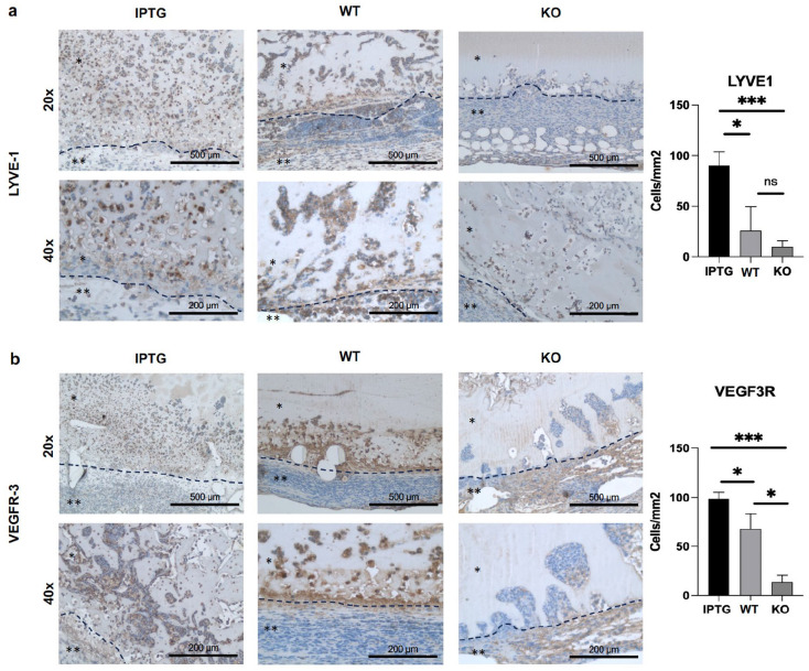
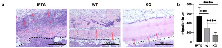
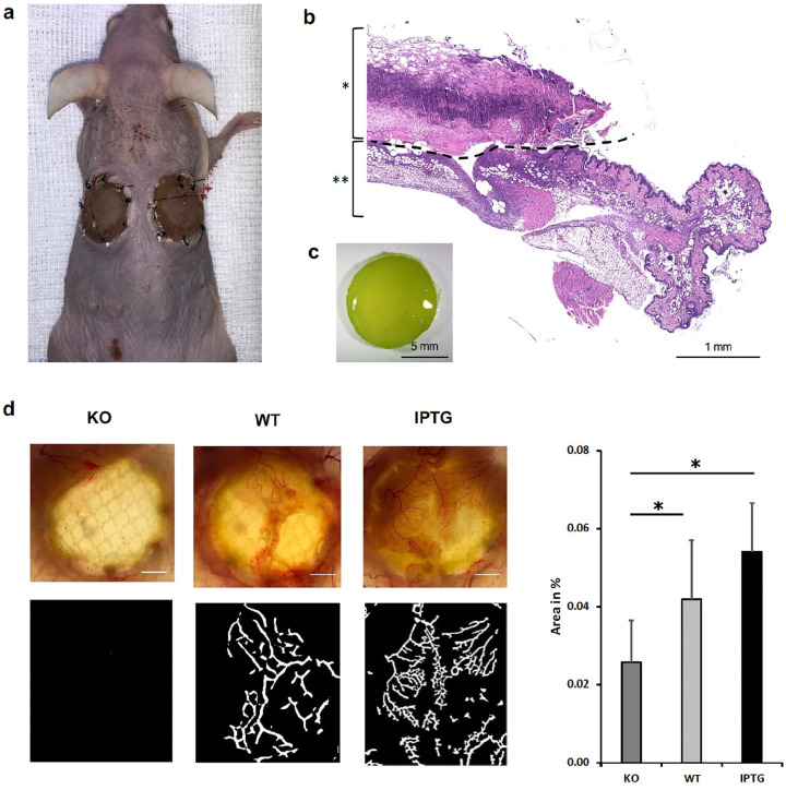
Chronic wounds represent an unresolved medical challenge with significant impact for patients' quality of life and global healthcare. Diverse in origin, ischemic-hypoxic and inflammatory conditions play central roles as pathological features that impede proper tissue regeneration. In this study, we propose an innovative approach to address this challenge. Our novel strategy utilizes photosynthetic biomaterials to restore the wound healing process firstly by promoting a normoxic, regeneration-supporting environment and secondly by mitigating inflammation through restoring lymphatic fluid transport and improving blood perfusion. We designed bioartificial scaffolds with photosynthetic cyanobacteria (Synechococcus sp. PCC 7002) and assessed their functional integration in a bilateral full-thickness skin defect on the backs of mice over a period of 7 days. Illuminated photosynthetic cyanobacteria facilitated local tissue oxygenation independent of blood perfusion. Additionally, genetic modification enabled the secretion of lymphangiogenic hyaluronic acid (HA) into the wound area. After 7 days, the scaffolds were explanted and histologically examined, assessing cell migration (HE staining) and protein expression (CD31, LYVE-1, VEGFR3, Ly6G, and F4/80). Results demonstrated successful colonization of bioartificial scaffolds with cyanobacteria. Following implantation into bilateral full-thickness skin defects, we observed an adherent vascularized basal layer beneath the bioactivated scaffolds after 7 days. Substantial increases in cell migration within bacteria-loaden scaffolds were noted, accompanied by a heightened expression of lymphatic (LYVE-1 and VEGFR3) and endothelial cell markers (CD31). Simultaneously, an augmented expression of acute (Ly6G) and late (F4/80) inflammatory proteins was observed. In summary, we developed a viable photosynthetic scaffold by integrating cyanobacteria into dermal regeneration materials (DRM), promoting the expression of lymphatic, endothelial, and inflammatory proteins under hypoxic conditions. The findings from this study represent a significant advancement in establishing autotrophic tissue engineering approaches, advocating for the use of photosynthetic cells in treating a broad spectrum of hypoxic conditions.
期刊介绍:
The Journal of Tissue Engineering (JTE) is a peer-reviewed, open-access journal dedicated to scientific research in the field of tissue engineering and its clinical applications. Our journal encompasses a wide range of interests, from the fundamental aspects of stem cells and progenitor cells, including their expansion to viable numbers, to an in-depth understanding of their differentiation processes. Join us in exploring the latest advancements in tissue engineering and its clinical translation.


 分享
分享
 求助内容:
求助内容: 应助结果提醒方式:
 扫码关注我们
