Felix Carbonell, Alex P Zijdenbos, Evan Hempel, Mihály Hajós, Barry J Bedell
{"title":"A novel method for harmonization of PET image spatial resolution without phantoms.","authors":"Felix Carbonell, Alex P Zijdenbos, Evan Hempel, Mihály Hajós, Barry J Bedell","doi":"10.1186/s40658-025-00740-9","DOIUrl":null,"url":null,"abstract":"<p><strong>Background: </strong>Estimation of the spatial resolution in real images is extremely important in several fields, including crystallography, optics, microscopy, and tomography. In human PET imaging, estimating spatial resolution typically involves the acquisition of images from a physical phantom, typically a Hoffman phantom, which poses a logistical burden, especially in large multi-center studies. Indeed, phantom images may not always be readily available, and this method requires constant monitoring of scanner updates or replacements, scanning protocol changes, and image reconstruction guidelines to establish a equivalence with scans acquired from human subjects.</p><p><strong>Methods: </strong>We propose a new computational approach that allows estimation of spatial resolution directly from human subject PET images. The proposed technique is based on the generalization of the logarithmic intensity plots in the 2D Fourier domain to the 3D case. The spatial resolution of the image is obtained through the estimated coefficients of a multiple linear regression problem having the logarithm of the squared norm of the Fourier transform as dependent variable and the squared 3D frequencies as multiple predictors.</p><p><strong>Results: </strong>The proposed approach was applied to a cohort of subjects consisting of [18F]florbetapir amyloid PET images and matching phantoms from a Phase II clinical trial, and a second cohort including β-amyloid, FDG, and tau PET images from the Alzheimer's Disease Neuroimaging Initiative (ADNI) study. The resulting in-plane and axial resolution estimators varied between 3.5 mm and 8.5 mm for both PET and matching phantom images. They also yielded less than one voxel size across-subjects variability in groups of images sharing the same PET scanner model and reconstruction parameters. For human PET images, we also proved that the spatial resolution estimators showed: (1) a very high reproducibility, as measured by intraclass correlation coefficients (ICC > 0.985), (2) a strong cross-tracer linear correlations, and (3) a high within-subject longitudinal consistency, as measured by the maximum difference value between pairs of visits from the same subject.</p><p><strong>Conclusions: </strong>Our novel approach does not only eliminate the need for surrogate phantom data, but also provides a general framework that can be applied to a wide range of tracers and other imaging modalities, such as SPECT.</p><p><strong>Clinical trial data: </strong>Cognito Therapeutics' OVERTURE clinical trial (NCT03556280, 2021-08-24), https://clinicaltrials.gov/study/NCT03556280 .</p>","PeriodicalId":11559,"journal":{"name":"EJNMMI Physics","volume":"12 1","pages":"23"},"PeriodicalIF":3.2000,"publicationDate":"2025-03-14","publicationTypes":"Journal Article","fieldsOfStudy":null,"isOpenAccess":false,"openAccessPdf":"https://www.ncbi.nlm.nih.gov/pmc/articles/PMC11906943/pdf/","citationCount":"0","resultStr":null,"platform":"Semanticscholar","paperid":null,"PeriodicalName":"EJNMMI Physics","FirstCategoryId":"3","ListUrlMain":"https://doi.org/10.1186/s40658-025-00740-9","RegionNum":2,"RegionCategory":"医学","ArticlePicture":[],"TitleCN":null,"AbstractTextCN":null,"PMCID":null,"EPubDate":"","PubModel":"","JCR":"Q2","JCRName":"RADIOLOGY, NUCLEAR MEDICINE & MEDICAL IMAGING","Score":null,"Total":0}
引用次数: 0
Abstract
Background: Estimation of the spatial resolution in real images is extremely important in several fields, including crystallography, optics, microscopy, and tomography. In human PET imaging, estimating spatial resolution typically involves the acquisition of images from a physical phantom, typically a Hoffman phantom, which poses a logistical burden, especially in large multi-center studies. Indeed, phantom images may not always be readily available, and this method requires constant monitoring of scanner updates or replacements, scanning protocol changes, and image reconstruction guidelines to establish a equivalence with scans acquired from human subjects.
Methods: We propose a new computational approach that allows estimation of spatial resolution directly from human subject PET images. The proposed technique is based on the generalization of the logarithmic intensity plots in the 2D Fourier domain to the 3D case. The spatial resolution of the image is obtained through the estimated coefficients of a multiple linear regression problem having the logarithm of the squared norm of the Fourier transform as dependent variable and the squared 3D frequencies as multiple predictors.
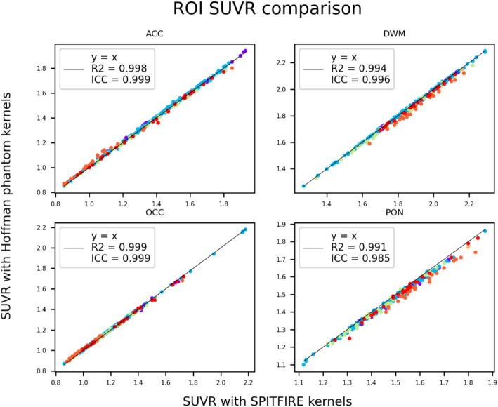
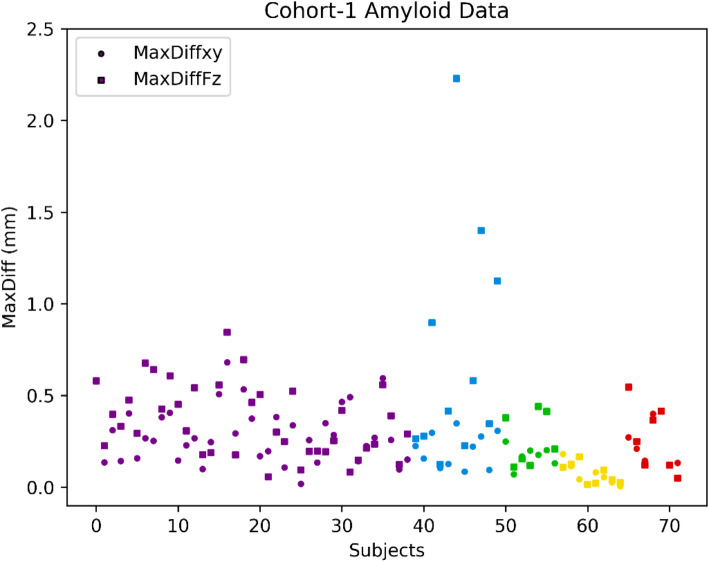
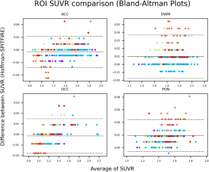
Results: The proposed approach was applied to a cohort of subjects consisting of [18F]florbetapir amyloid PET images and matching phantoms from a Phase II clinical trial, and a second cohort including β-amyloid, FDG, and tau PET images from the Alzheimer's Disease Neuroimaging Initiative (ADNI) study. The resulting in-plane and axial resolution estimators varied between 3.5 mm and 8.5 mm for both PET and matching phantom images. They also yielded less than one voxel size across-subjects variability in groups of images sharing the same PET scanner model and reconstruction parameters. For human PET images, we also proved that the spatial resolution estimators showed: (1) a very high reproducibility, as measured by intraclass correlation coefficients (ICC > 0.985), (2) a strong cross-tracer linear correlations, and (3) a high within-subject longitudinal consistency, as measured by the maximum difference value between pairs of visits from the same subject.
Conclusions: Our novel approach does not only eliminate the need for surrogate phantom data, but also provides a general framework that can be applied to a wide range of tracers and other imaging modalities, such as SPECT.
期刊介绍:
EJNMMI Physics is an international platform for scientists, users and adopters of nuclear medicine with a particular interest in physics matters. As a companion journal to the European Journal of Nuclear Medicine and Molecular Imaging, this journal has a multi-disciplinary approach and welcomes original materials and studies with a focus on applied physics and mathematics as well as imaging systems engineering and prototyping in nuclear medicine. This includes physics-driven approaches or algorithms supported by physics that foster early clinical adoption of nuclear medicine imaging and therapy.


 分享
分享
 求助内容:
求助内容: 应助结果提醒方式:
 扫码关注我们
