Daniel Henrique Koga, Marcos Martins Curi, Joel Ferreira Santiago Junior, Aldieris Alves Pesqueira, Wagner José Sousa Carvalho, Márcio Campaner, Camila Lopes Cardoso
{"title":"Pterygoid implant: extensometric and photoelastic analysis of a maxillary rehabilitation model.","authors":"Daniel Henrique Koga, Marcos Martins Curi, Joel Ferreira Santiago Junior, Aldieris Alves Pesqueira, Wagner José Sousa Carvalho, Márcio Campaner, Camila Lopes Cardoso","doi":"10.1590/1807-3107bor-2025.vol39.030","DOIUrl":null,"url":null,"abstract":"<p><p>Pterygoid implants have been demonstrated to have a high success rate. Nevertheless, there are few biomechanical tests to evaluate the tensile forces induced by force dissipation in peri-implant tissues. This study employed photoelasticity and extensometry to demonstrate and compare the biomechanical behavior of non-axial implants in a pterygoid model and a conventional model of oral rehabilitation, thus allowing for qualitative and quantitative analysis. Two models received an implant measuring 3.75 x 13 mm in the canine pillar at a 90 ° angle to the Frankfurt plane. In the control group, an implant with a diameter of 3.75 mm and a length of 11 mm was placed in the maxillary tuberosity parallel the medial implant. In the study group, an implant with a diameter of 3.75 mm and a length of 11 mm was installed with an angulation of 45 degrees in the antero-posterior direction and 15 degrees in the buccal-palatal direction, with apical anchorage in the pterygoid process of the sphenoid bone. In the extensometric analysis, the models were subjected to five cycles of repeated axial tensile loading (100 N) at a rate of 0.5 mm/min. A computer was connected to the amplifier in order to record the output signal of the polyurethane surface, and the acquisition system software was employed to record the data. The data were analyzed in accordance with data distribution, as determined by the Shapiro-Wilk test and equality of variance. Subsequently, the data were classified according to the variables. The Student's t-test was employed when normal distribution of variances was identified, whereas the Mann-Whitney U test was utilized for data with non-normal distribution. A 5% significance level was employed. In the photoelastic analysis, replicas of both configurations were produced using photoelastic resin. The models were subjected to a single axial loading cycle, with a load of 100 N applied at a rate of 0.5 mm/min, and the resulting stress was observed under a circular polariscope. Photographs were taken at two time points: before and after loading. These images were then processed by the same operator using a computer graphics program, allowing for a more straightforward analysis of stress distribution. This was achieved by the formation of isochromatic fringes. The results of the strain gauge analysis revealed no statistically significant differences between the two groups (p = 0.37) or between the anterior (p = 0.08) and posterior (p = 0.74) implants. The photoelasticity analysis revealed the presence of high-intensity isochromatic fringes at the apex of the axial implant in the control model, as well as in the cervical-distal and apical regions of the pterygoid implant, where a high concentration was also observed. Although no statistically significant results were obtained from the quantitative analysis, our findings suggest that the favorable outcomes observed in the clinical studies are due to the high force dissipation observed in the pterygoid plate, which is composed of dense cortical bone.</p>","PeriodicalId":9240,"journal":{"name":"Brazilian oral research","volume":"39 ","pages":"e030"},"PeriodicalIF":1.3000,"publicationDate":"2025-03-10","publicationTypes":"Journal Article","fieldsOfStudy":null,"isOpenAccess":false,"openAccessPdf":"https://www.ncbi.nlm.nih.gov/pmc/articles/PMC11893003/pdf/","citationCount":"0","resultStr":null,"platform":"Semanticscholar","paperid":null,"PeriodicalName":"Brazilian oral research","FirstCategoryId":"3","ListUrlMain":"https://doi.org/10.1590/1807-3107bor-2025.vol39.030","RegionNum":4,"RegionCategory":"医学","ArticlePicture":[],"TitleCN":null,"AbstractTextCN":null,"PMCID":null,"EPubDate":"2025/1/1 0:00:00","PubModel":"eCollection","JCR":"Q3","JCRName":"DENTISTRY, ORAL SURGERY & MEDICINE","Score":null,"Total":0}
引用次数: 0
Abstract
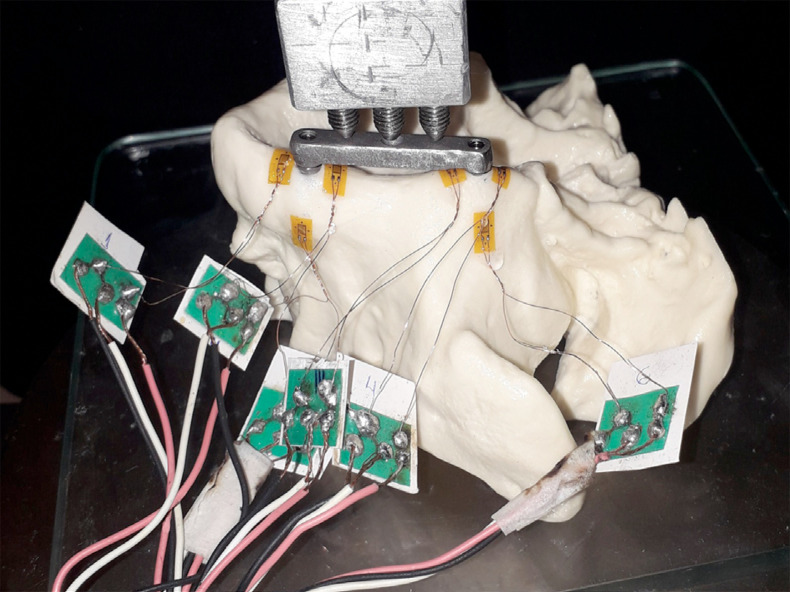
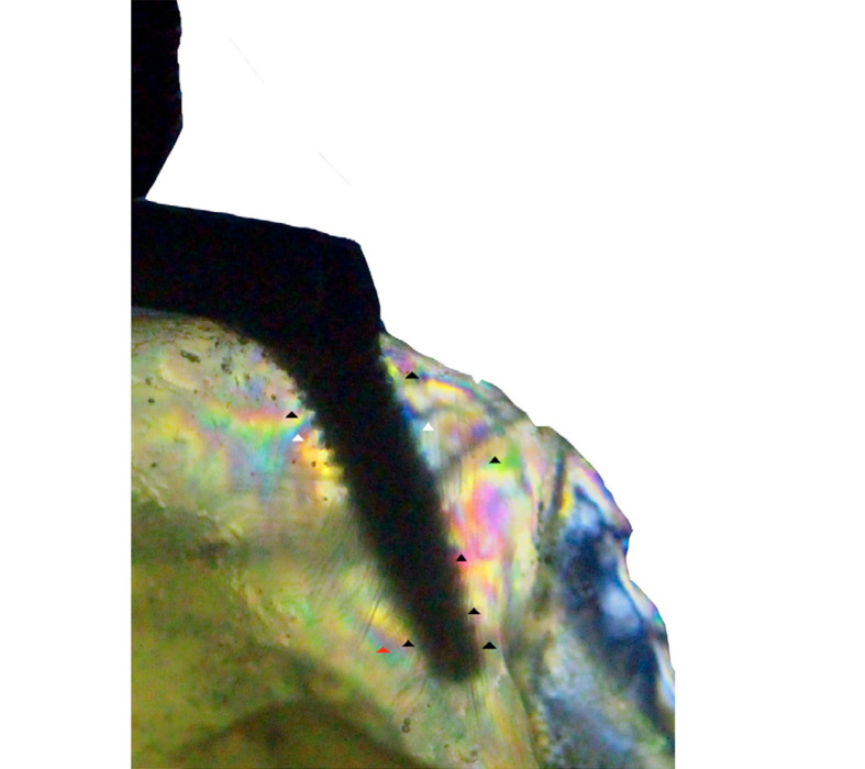
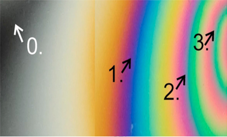
Pterygoid implants have been demonstrated to have a high success rate. Nevertheless, there are few biomechanical tests to evaluate the tensile forces induced by force dissipation in peri-implant tissues. This study employed photoelasticity and extensometry to demonstrate and compare the biomechanical behavior of non-axial implants in a pterygoid model and a conventional model of oral rehabilitation, thus allowing for qualitative and quantitative analysis. Two models received an implant measuring 3.75 x 13 mm in the canine pillar at a 90 ° angle to the Frankfurt plane. In the control group, an implant with a diameter of 3.75 mm and a length of 11 mm was placed in the maxillary tuberosity parallel the medial implant. In the study group, an implant with a diameter of 3.75 mm and a length of 11 mm was installed with an angulation of 45 degrees in the antero-posterior direction and 15 degrees in the buccal-palatal direction, with apical anchorage in the pterygoid process of the sphenoid bone. In the extensometric analysis, the models were subjected to five cycles of repeated axial tensile loading (100 N) at a rate of 0.5 mm/min. A computer was connected to the amplifier in order to record the output signal of the polyurethane surface, and the acquisition system software was employed to record the data. The data were analyzed in accordance with data distribution, as determined by the Shapiro-Wilk test and equality of variance. Subsequently, the data were classified according to the variables. The Student's t-test was employed when normal distribution of variances was identified, whereas the Mann-Whitney U test was utilized for data with non-normal distribution. A 5% significance level was employed. In the photoelastic analysis, replicas of both configurations were produced using photoelastic resin. The models were subjected to a single axial loading cycle, with a load of 100 N applied at a rate of 0.5 mm/min, and the resulting stress was observed under a circular polariscope. Photographs were taken at two time points: before and after loading. These images were then processed by the same operator using a computer graphics program, allowing for a more straightforward analysis of stress distribution. This was achieved by the formation of isochromatic fringes. The results of the strain gauge analysis revealed no statistically significant differences between the two groups (p = 0.37) or between the anterior (p = 0.08) and posterior (p = 0.74) implants. The photoelasticity analysis revealed the presence of high-intensity isochromatic fringes at the apex of the axial implant in the control model, as well as in the cervical-distal and apical regions of the pterygoid implant, where a high concentration was also observed. Although no statistically significant results were obtained from the quantitative analysis, our findings suggest that the favorable outcomes observed in the clinical studies are due to the high force dissipation observed in the pterygoid plate, which is composed of dense cortical bone.


 分享
分享
 求助内容:
求助内容: 应助结果提醒方式:
 扫码关注我们
