{"title":"Effects of breast structure on high-intensity focused ultrasound focal error.","authors":"Kohei Okita, Ryuta Narumi, Takashi Azuma, Hidemi Furusawa, Junichi Shidooka, Shu Takagi, Yoichiro Matsumoto","doi":"10.1186/s40349-018-0111-9","DOIUrl":null,"url":null,"abstract":"<p><strong>Background: </strong>The development of imaging technologies and breast cancer screening allowed early detection of breast cancers. High-intensity focused ultrasound (HIFU) is a non-invasive cancer treatment, but the success of HIFU ablation was depending on the system type, imaging technique, ablation protocol, and patient selection. Therefore, we aimed to determine the relationship between breast tissue structure and focal error during breast cancer HIFU treatment.</p><p><strong>Methods: </strong>Numerical simulations of the breast cancer HIFU ablation were performed using digital breast phantoms constructed using the magnetic resonance imaging data obtained from 12 patients.</p><p><strong>Results: </strong>The focal shapes were distorted despite breast tissue representing soft tissue. Focal errors are caused by the complex distribution of fibroglandular tissue, and they depend on the target position and the arrangement of the transducer. We demonstrated that the focusing ratio increases with the decrease in the local acoustic inhomogeneity, implying that it may be used as an indicator to reduce the HIFU focal error depending on the breast structure.</p><p><strong>Conclusions: </strong>The obtained results demonstrated that the focal error observed during the breast cancer HIFU treatment is highly dependent on the structure of fibroglandular tissue. The optimal arrangement of the transducer to the target can be obtained by minimizing the local acoustic inhomogeneity before the breast cancer HIFU treatment.</p>","PeriodicalId":90245,"journal":{"name":"Journal of therapeutic ultrasound","volume":"6 ","pages":"4"},"PeriodicalIF":0.0000,"publicationDate":"2018-06-20","publicationTypes":"Journal Article","fieldsOfStudy":null,"isOpenAccess":false,"openAccessPdf":"https://sci-hub-pdf.com/10.1186/s40349-018-0111-9","citationCount":"10","resultStr":null,"platform":"Semanticscholar","paperid":null,"PeriodicalName":"Journal of therapeutic ultrasound","FirstCategoryId":"1085","ListUrlMain":"https://doi.org/10.1186/s40349-018-0111-9","RegionNum":0,"RegionCategory":null,"ArticlePicture":[],"TitleCN":null,"AbstractTextCN":null,"PMCID":null,"EPubDate":"2018/1/1 0:00:00","PubModel":"eCollection","JCR":"","JCRName":"","Score":null,"Total":0}
引用次数: 10
Abstract
Background: The development of imaging technologies and breast cancer screening allowed early detection of breast cancers. High-intensity focused ultrasound (HIFU) is a non-invasive cancer treatment, but the success of HIFU ablation was depending on the system type, imaging technique, ablation protocol, and patient selection. Therefore, we aimed to determine the relationship between breast tissue structure and focal error during breast cancer HIFU treatment.
Methods: Numerical simulations of the breast cancer HIFU ablation were performed using digital breast phantoms constructed using the magnetic resonance imaging data obtained from 12 patients.
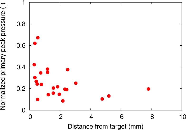
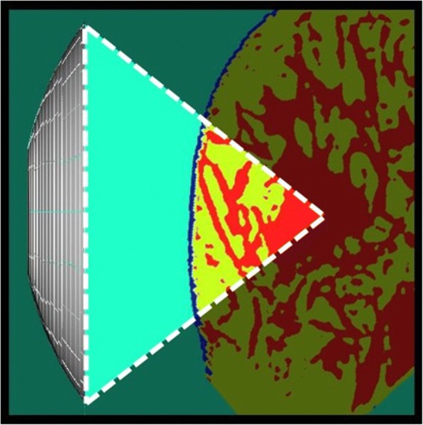
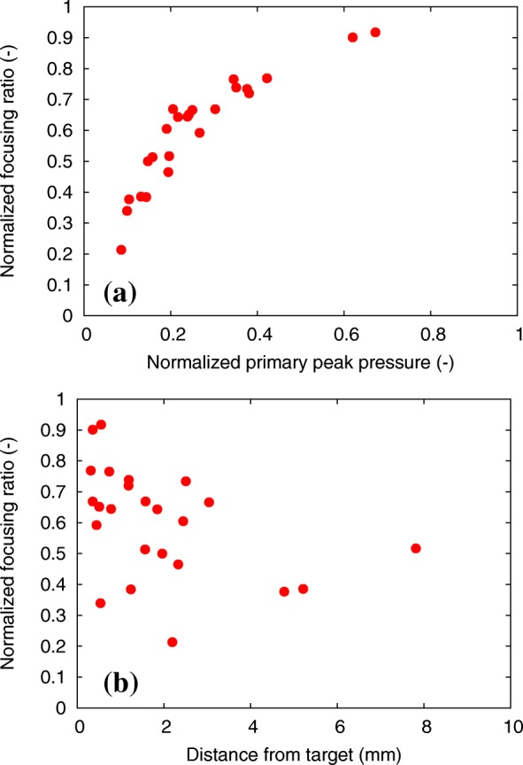
Results: The focal shapes were distorted despite breast tissue representing soft tissue. Focal errors are caused by the complex distribution of fibroglandular tissue, and they depend on the target position and the arrangement of the transducer. We demonstrated that the focusing ratio increases with the decrease in the local acoustic inhomogeneity, implying that it may be used as an indicator to reduce the HIFU focal error depending on the breast structure.
Conclusions: The obtained results demonstrated that the focal error observed during the breast cancer HIFU treatment is highly dependent on the structure of fibroglandular tissue. The optimal arrangement of the transducer to the target can be obtained by minimizing the local acoustic inhomogeneity before the breast cancer HIFU treatment.


 分享
分享

 求助内容:
求助内容: 应助结果提醒方式:
 扫码关注我们
