Yong-Jik Lee, Hyun Soo Kim, Hong Seog Seo, Jin Oh Na, You-Na Jang, Yoon-Mi Han, Hyun-Min Kim
{"title":"Stimulation of Alpha<sub>1</sub>-Adrenergic Receptor Ameliorates Cellular Functions of Multiorgans beyond Vasomotion through PPAR<i>δ</i>.","authors":"Yong-Jik Lee, Hyun Soo Kim, Hong Seog Seo, Jin Oh Na, You-Na Jang, Yoon-Mi Han, Hyun-Min Kim","doi":"10.1155/2020/3785137","DOIUrl":null,"url":null,"abstract":"<p><p>Cells can shift their metabolism between glycolysis and oxidative phosphorylation to enact their cell fate program in response to external signals. Widely distributed <i>α</i> <sub>1</sub>-adrenergic receptors (ARs) are physiologically stimulated during exercise, were reported to associate with the activating energetic AMPK pathway, and are expected to have biological effects beyond their hemodynamic effects. To investigate the effects and mechanism of AR stimulation on the physiology of the whole body, various <i>in vitro</i> and <i>in vivo</i> experiments were conducted using the AR agonist midodrine, 2-amino-<i>N</i>-[2-(2,5-dimethoxyphenyl)-2-hydroxy-ethyl]-acetamide. The expression of various biomarkers involved in ATP production was estimated through Western blotting, reverse transcription polymerase chain reaction, oxygen consumption rate, enzyme-linked immunosorbent assay (ELISA), fluorescence staining, and Oil red O staining in several cell lines (skeletal muscle, cardiac muscle, liver, macrophage, vascular endothelial, and adipose cells). In spontaneously hypertensive rats, blood pressure, blood analysis, organ-specific biomarkers, and general biomolecules related to ATP production were measured with Western blot analysis, immunohistochemistry, ELISA, and echocardiography. Pharmacological activation of <i>α</i> <sub>1</sub>-adrenergic receptors in C2C12 skeletal muscle cells promoted mitochondrial oxidative phosphorylation and ATP production by increasing the expression of catabolic molecules, including PPAR<i>δ</i>, AMPK, and PGC-1<i>α</i>, through cytosolic calcium signaling and increased GLUT4 expression, as seen in exercise. It also activated those energetic molecules and mitochondrial oxidative phosphorylation with cardiomyocytes, endothelial cells, adipocytes, macrophages, and hepatic cells and affected their relevant cell-specific biological functions. All of those effects occurred around 3 h (and peaked 6 h) after midodrine treatment. In spontaneously hypertensive rats, <i>α</i> <sub>1</sub>-adrenergic receptor stimulation affected mitochondrial oxidative phosphorylation and ATP production by activating PPAR<i>δ</i>, AMPK, and PGC-1<i>α</i> and the relevant biologic functions of multiple organs, suggesting organ crosstalk. The treatment lowered blood pressure, fat and body weight, cholesterol levels, and inflammatory activity; increased ATP content and insulin sensitivity in skeletal muscles; and increased cardiac contractile function without exercise training. These results suggest that the activation of <i>α</i> <sub>1</sub>-adrenergic receptor stimulates energetic reprogramming via PPAR<i>δ</i> that increases mitochondrial oxidative phosphorylation and has healthy and organ-specific biological effects in multiple organs, including skeletal muscle, beyond its vasomotion effect. In addition, the action mechanism of <i>α</i> <sub>1</sub>-adrenergic receptor may be mainly exerted via PPAR<i>δ</i>.</p>","PeriodicalId":20439,"journal":{"name":"PPAR Research","volume":"2020 ","pages":"3785137"},"PeriodicalIF":3.1000,"publicationDate":"2020-02-01","publicationTypes":"Journal Article","fieldsOfStudy":null,"isOpenAccess":false,"openAccessPdf":"https://sci-hub-pdf.com/10.1155/2020/3785137","citationCount":"10","resultStr":null,"platform":"Semanticscholar","paperid":null,"PeriodicalName":"PPAR Research","FirstCategoryId":"3","ListUrlMain":"https://doi.org/10.1155/2020/3785137","RegionNum":3,"RegionCategory":"医学","ArticlePicture":[],"TitleCN":null,"AbstractTextCN":null,"PMCID":null,"EPubDate":"2020/1/1 0:00:00","PubModel":"eCollection","JCR":"Q2","JCRName":"MEDICINE, RESEARCH & EXPERIMENTAL","Score":null,"Total":0}
引用次数: 10
Abstract
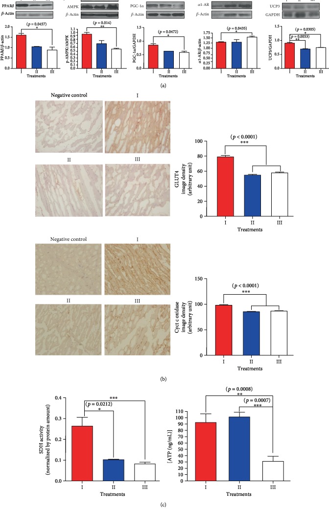
Cells can shift their metabolism between glycolysis and oxidative phosphorylation to enact their cell fate program in response to external signals. Widely distributed α1-adrenergic receptors (ARs) are physiologically stimulated during exercise, were reported to associate with the activating energetic AMPK pathway, and are expected to have biological effects beyond their hemodynamic effects. To investigate the effects and mechanism of AR stimulation on the physiology of the whole body, various in vitro and in vivo experiments were conducted using the AR agonist midodrine, 2-amino-N-[2-(2,5-dimethoxyphenyl)-2-hydroxy-ethyl]-acetamide. The expression of various biomarkers involved in ATP production was estimated through Western blotting, reverse transcription polymerase chain reaction, oxygen consumption rate, enzyme-linked immunosorbent assay (ELISA), fluorescence staining, and Oil red O staining in several cell lines (skeletal muscle, cardiac muscle, liver, macrophage, vascular endothelial, and adipose cells). In spontaneously hypertensive rats, blood pressure, blood analysis, organ-specific biomarkers, and general biomolecules related to ATP production were measured with Western blot analysis, immunohistochemistry, ELISA, and echocardiography. Pharmacological activation of α1-adrenergic receptors in C2C12 skeletal muscle cells promoted mitochondrial oxidative phosphorylation and ATP production by increasing the expression of catabolic molecules, including PPARδ, AMPK, and PGC-1α, through cytosolic calcium signaling and increased GLUT4 expression, as seen in exercise. It also activated those energetic molecules and mitochondrial oxidative phosphorylation with cardiomyocytes, endothelial cells, adipocytes, macrophages, and hepatic cells and affected their relevant cell-specific biological functions. All of those effects occurred around 3 h (and peaked 6 h) after midodrine treatment. In spontaneously hypertensive rats, α1-adrenergic receptor stimulation affected mitochondrial oxidative phosphorylation and ATP production by activating PPARδ, AMPK, and PGC-1α and the relevant biologic functions of multiple organs, suggesting organ crosstalk. The treatment lowered blood pressure, fat and body weight, cholesterol levels, and inflammatory activity; increased ATP content and insulin sensitivity in skeletal muscles; and increased cardiac contractile function without exercise training. These results suggest that the activation of α1-adrenergic receptor stimulates energetic reprogramming via PPARδ that increases mitochondrial oxidative phosphorylation and has healthy and organ-specific biological effects in multiple organs, including skeletal muscle, beyond its vasomotion effect. In addition, the action mechanism of α1-adrenergic receptor may be mainly exerted via PPARδ.
期刊介绍:
PPAR Research is a peer-reviewed, Open Access journal that publishes original research and review articles on advances in basic research focusing on mechanisms involved in the activation of peroxisome proliferator-activated receptors (PPARs), as well as their role in the regulation of cellular differentiation, development, energy homeostasis and metabolic function. The journal also welcomes preclinical and clinical trials of drugs that can modulate PPAR activity, with a view to treating chronic diseases and disorders such as dyslipidemia, diabetes, adipocyte differentiation, inflammation, cancer, lung diseases, neurodegenerative disorders, and obesity.


 分享
分享
 求助内容:
求助内容: 应助结果提醒方式:
 扫码关注我们
