Ian Grierson, Don Minckler, Marian K Rippy, Andrew J Marshall, Nathalie Collignon, Jessica Bianco, Benoit Detry, Murray A Johnstone
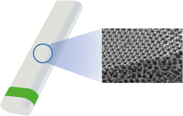
{"title":"A novel suprachoroidal microinvasive glaucoma implant: in vivo biocompatibility and biointegration.","authors":"Ian Grierson, Don Minckler, Marian K Rippy, Andrew J Marshall, Nathalie Collignon, Jessica Bianco, Benoit Detry, Murray A Johnstone","doi":"10.1186/s42490-020-00045-1","DOIUrl":null,"url":null,"abstract":"<p><strong>Background: </strong>A major challenge for any glaucoma implant is their ability to provide long-term intraocular pressure lowering efficacy. The formation of a low-permeability fibrous capsule around the device often leads to obstructed drainage channels, which may impair the drainage function of devices. These foreign body-related limitations point to the need to develop biologically inert biomaterials to improve performance in reaching long-term intraocular pressure reduction. The aim of this study was to evaluate in vivo (in rabbits) the ocular biocompatibility and tissue integration of a novel suprachoroidal microinvasive glaucoma implant, MINIject™ (iSTAR Medical, Wavre, Belgium).</p><p><strong>Results: </strong>In two rabbit studies, no biocompatibility issue was induced by the suprachoroidal, ab-externo implantation of the MINIject™ device. Clinical evaluation throughout the 6 post-operative months between the sham and test groups were similar, suggesting most reactions were related to the ab-externo surgical technique used for rabbits, rather than the implant material itself. Histological analysis of ocular tissues at post-operative months 1, 3 and 6 revealed that the implant was well-tolerated and induced only minimal fibroplasia and thus minimal encapsulation around the implant. The microporous structure of the device became rapidly colonized by cells, mostly by macrophages through cell migration, which do not, by their nature, impede the flow of aqueous humor through the device. Time-course analysis showed that once established, pore colonization was stable over time. No fibrosis nor dense connective tissue development were observed within any implant at any time point. The presence of pore colonization may be the process by which encapsulation around the implant is minimized, thus preserving the permeability of the surrounding tissues. No degradation nor structural changes of the implant occurred during the course of both studies.</p><p><strong>Conclusions: </strong>The novel MINIject™ microinvasive glaucoma implant was well-tolerated in ocular tissues of rabbits, with observance of biointegration, and no biocompatibility issues. Minimal fibrous encapsulation and stable cellular pore colonization provided evidence of preserved drainage properties over time, suggesting that the implant may produce a long-term ability to enhance aqueous outflow.</p>","PeriodicalId":72425,"journal":{"name":"BMC biomedical engineering","volume":"2 ","pages":"10"},"PeriodicalIF":0.0000,"publicationDate":"2020-10-14","publicationTypes":"Journal Article","fieldsOfStudy":null,"isOpenAccess":false,"openAccessPdf":"https://sci-hub-pdf.com/10.1186/s42490-020-00045-1","citationCount":"10","resultStr":null,"platform":"Semanticscholar","paperid":null,"PeriodicalName":"BMC biomedical engineering","FirstCategoryId":"1085","ListUrlMain":"https://doi.org/10.1186/s42490-020-00045-1","RegionNum":0,"RegionCategory":null,"ArticlePicture":[],"TitleCN":null,"AbstractTextCN":null,"PMCID":null,"EPubDate":"2020/1/1 0:00:00","PubModel":"eCollection","JCR":"","JCRName":"","Score":null,"Total":0}
引用次数: 10
Abstract
Background: A major challenge for any glaucoma implant is their ability to provide long-term intraocular pressure lowering efficacy. The formation of a low-permeability fibrous capsule around the device often leads to obstructed drainage channels, which may impair the drainage function of devices. These foreign body-related limitations point to the need to develop biologically inert biomaterials to improve performance in reaching long-term intraocular pressure reduction. The aim of this study was to evaluate in vivo (in rabbits) the ocular biocompatibility and tissue integration of a novel suprachoroidal microinvasive glaucoma implant, MINIject™ (iSTAR Medical, Wavre, Belgium).
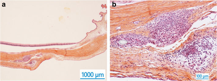
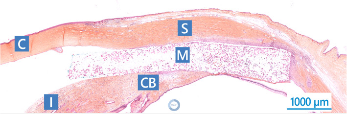
Results: In two rabbit studies, no biocompatibility issue was induced by the suprachoroidal, ab-externo implantation of the MINIject™ device. Clinical evaluation throughout the 6 post-operative months between the sham and test groups were similar, suggesting most reactions were related to the ab-externo surgical technique used for rabbits, rather than the implant material itself. Histological analysis of ocular tissues at post-operative months 1, 3 and 6 revealed that the implant was well-tolerated and induced only minimal fibroplasia and thus minimal encapsulation around the implant. The microporous structure of the device became rapidly colonized by cells, mostly by macrophages through cell migration, which do not, by their nature, impede the flow of aqueous humor through the device. Time-course analysis showed that once established, pore colonization was stable over time. No fibrosis nor dense connective tissue development were observed within any implant at any time point. The presence of pore colonization may be the process by which encapsulation around the implant is minimized, thus preserving the permeability of the surrounding tissues. No degradation nor structural changes of the implant occurred during the course of both studies.
Conclusions: The novel MINIject™ microinvasive glaucoma implant was well-tolerated in ocular tissues of rabbits, with observance of biointegration, and no biocompatibility issues. Minimal fibrous encapsulation and stable cellular pore colonization provided evidence of preserved drainage properties over time, suggesting that the implant may produce a long-term ability to enhance aqueous outflow.


 分享
分享

 求助内容:
求助内容: 应助结果提醒方式:
 扫码关注我们
