{"title":"Live-cell imaging probes to track chromatin modification dynamics","authors":"Yuko Sato;Masaru Nakao;Hiroshi Kimura","doi":"10.1093/jmicro/dfab030","DOIUrl":null,"url":null,"abstract":"The spatiotemporal organization of chromatin is regulated at different levels in the nucleus. Epigenetic modifications such as DNA methylation and histone modifications are involved in chromatin regulation and play fundamental roles in genome function. While the one-dimensional epigenomic landscape in many cell types has been revealed by chromatin immunoprecipitation and sequencing, the dynamic changes of chromatin modifications and their relevance to chromatin organization and genome function remain elusive. Live-cell probes to visualize chromatin and its modifications have become powerful tools to monitor dynamic chromatin regulation. Bulk chromatin can be visualized by both small fluorescent dyes and fluorescent proteins, and specific endogenous genomic loci have been detected by adapting genome-editing tools. To track chromatin modifications in living cells, various types of probes have been developed. Protein domains that bind weakly to specific modifications, such as chromodomains for histone methylation, can be repeated to create a tighter binding probe that can then be tagged with a fluorescent protein. It has also been demonstrated that antigen-binding fragments and single-chain variable fragments from modification-specific antibodies can serve as binding probes without disturbing cell division, development and differentiation. These modification-binding modules are used in modification sensors based on fluorescence/Förster resonance energy transfer to measure the intramolecular conformational changes triggered by modifications. Other probes can be created using a bivalent binding system, such as fluorescence complementation or luciferase chemiluminescence. Live-cell chromatin modification imaging using these probes will address dynamic chromatin regulation and will be useful for assaying and screening effective epigenome drugs in cells and organisms.","PeriodicalId":18515,"journal":{"name":"Microscopy","volume":"70 5","pages":"415-422"},"PeriodicalIF":1.8000,"publicationDate":"2021-10-01","publicationTypes":"Journal Article","fieldsOfStudy":null,"isOpenAccess":false,"openAccessPdf":"https://ieeexplore.ieee.org/stamp/stamp.jsp?tp=&arnumber=9623672","citationCount":"8","resultStr":null,"platform":"Semanticscholar","paperid":null,"PeriodicalName":"Microscopy","FirstCategoryId":"5","ListUrlMain":"https://ieeexplore.ieee.org/document/9623672/","RegionNum":4,"RegionCategory":"工程技术","ArticlePicture":[],"TitleCN":null,"AbstractTextCN":null,"PMCID":null,"EPubDate":"","PubModel":"","JCR":"","JCRName":"","Score":null,"Total":0}
引用次数: 8
Abstract
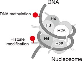
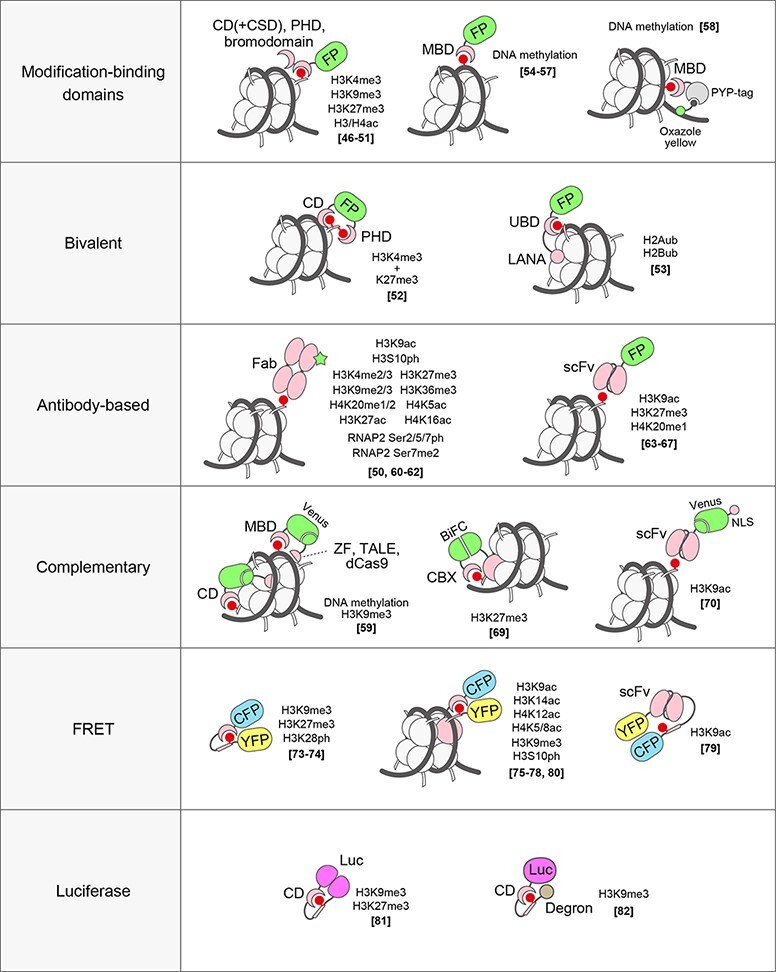
The spatiotemporal organization of chromatin is regulated at different levels in the nucleus. Epigenetic modifications such as DNA methylation and histone modifications are involved in chromatin regulation and play fundamental roles in genome function. While the one-dimensional epigenomic landscape in many cell types has been revealed by chromatin immunoprecipitation and sequencing, the dynamic changes of chromatin modifications and their relevance to chromatin organization and genome function remain elusive. Live-cell probes to visualize chromatin and its modifications have become powerful tools to monitor dynamic chromatin regulation. Bulk chromatin can be visualized by both small fluorescent dyes and fluorescent proteins, and specific endogenous genomic loci have been detected by adapting genome-editing tools. To track chromatin modifications in living cells, various types of probes have been developed. Protein domains that bind weakly to specific modifications, such as chromodomains for histone methylation, can be repeated to create a tighter binding probe that can then be tagged with a fluorescent protein. It has also been demonstrated that antigen-binding fragments and single-chain variable fragments from modification-specific antibodies can serve as binding probes without disturbing cell division, development and differentiation. These modification-binding modules are used in modification sensors based on fluorescence/Förster resonance energy transfer to measure the intramolecular conformational changes triggered by modifications. Other probes can be created using a bivalent binding system, such as fluorescence complementation or luciferase chemiluminescence. Live-cell chromatin modification imaging using these probes will address dynamic chromatin regulation and will be useful for assaying and screening effective epigenome drugs in cells and organisms.
期刊介绍:
Microscopy, previously Journal of Electron Microscopy, promotes research combined with any type of microscopy techniques, applied in life and material sciences. Microscopy is the official journal of the Japanese Society of Microscopy.


 分享
分享
 求助内容:
求助内容: 应助结果提醒方式:
 扫码关注我们
