{"title":"Nobiletin Attenuates Pathological Cardiac Remodeling after Myocardial Infarction via Activating PPAR<i>γ</i> and PGC1<i>α</i>.","authors":"Yufei Zhou, Ting Yin, Mengsha Shi, Mengli Chen, Xiaodong Wu, Kai Wang, Iokfai Cheang, Yanxiu Li, Hongcai Shang, Haifeng Zhang, Xinli Li","doi":"10.1155/2021/9947656","DOIUrl":null,"url":null,"abstract":"<p><strong>Materials and methods: </strong>C57BL/6 mice were treated with coronary artery ligation to generate an MI model, followed by treatment for 3 weeks with NOB (50 mg/kg/d) or vehicle (50 mg/kg/d), with or without the peroxisome proliferator-activated receptor gamma (PPAR<i>γ</i>) inhibitor T0070907 (1 mg/kg/d). Cardiac function (echocardiography, survival rate, Evans blue, and triphenyl tetrazolium chloride staining), fibrosis (Masson's trichrome staining, quantitative real-time polymerase chain reaction (qRT-PCR), and western blot (WB)), hypertrophy (haematoxylin-eosin staining, wheat germ agglutinin staining, and qRT-PCR), and apoptosis (WB and terminal deoxynucleotidyl transferase dUTP nick-end labelling (TUNEL) staining) were evaluated. Hypoxia-induced apoptosis (TUNEL, WB) and phenylephrine- (PE-) induced pathological hypertrophy (immunofluorescence staining, qRT-PCR) models were established in primary neonatal rat ventricular myocytes (NRVMs). The effects of NOB with or without T0070907 were examined for the expression of PPAR<i>γ</i> and PPAR<i>γ</i> coactivator 1<i>α</i> (PGC1<i>α</i>) by WB in mice and NRVMs. The potential downstream effectors of PPAR<i>γ</i> were further analyzed by WB in mice.</p><p><strong>Results: </strong>Following MI in mice, NOB intervention enhanced cardiac function across three predominant dimensions of pathological cardiac remodeling, which reflected in decreasing cardiac fibrosis, apoptosis, and hypertrophy decompensation. NOB intervention also alleviated apoptosis and hypertrophy in NRVMs. NOB intervention upregulated PPAR<i>γ</i> and PGC1<i>α in vivo</i> and <i>in vitro</i>. Furthermore, the PPAR<i>γ</i> inhibitor abolished the protective effects of NOB against pathological cardiac remodeling during the progression from MI to CHF. The potential downstream effectors of PPAR<i>γ</i> were nuclear factor erythroid 2-related factor 2 (Nrf-2) and heme oxygenase 1 (HO-1).</p><p><strong>Conclusions: </strong>Our findings suggested that NOB alleviates pathological cardiac remodeling after MI via PPAR<i>γ</i> and PGC1<i>α</i> upregulation.</p>","PeriodicalId":20439,"journal":{"name":"PPAR Research","volume":" ","pages":"9947656"},"PeriodicalIF":3.1000,"publicationDate":"2021-08-06","publicationTypes":"Journal Article","fieldsOfStudy":null,"isOpenAccess":false,"openAccessPdf":"https://www.ncbi.nlm.nih.gov/pmc/articles/PMC8373512/pdf/","citationCount":"9","resultStr":null,"platform":"Semanticscholar","paperid":null,"PeriodicalName":"PPAR Research","FirstCategoryId":"3","ListUrlMain":"https://doi.org/10.1155/2021/9947656","RegionNum":3,"RegionCategory":"医学","ArticlePicture":[],"TitleCN":null,"AbstractTextCN":null,"PMCID":null,"EPubDate":"2021/1/1 0:00:00","PubModel":"eCollection","JCR":"Q2","JCRName":"MEDICINE, RESEARCH & EXPERIMENTAL","Score":null,"Total":0}
引用次数: 9
Abstract
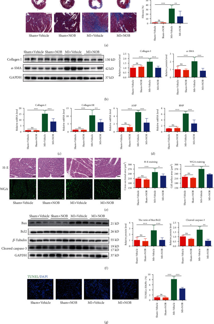
Materials and methods: C57BL/6 mice were treated with coronary artery ligation to generate an MI model, followed by treatment for 3 weeks with NOB (50 mg/kg/d) or vehicle (50 mg/kg/d), with or without the peroxisome proliferator-activated receptor gamma (PPARγ) inhibitor T0070907 (1 mg/kg/d). Cardiac function (echocardiography, survival rate, Evans blue, and triphenyl tetrazolium chloride staining), fibrosis (Masson's trichrome staining, quantitative real-time polymerase chain reaction (qRT-PCR), and western blot (WB)), hypertrophy (haematoxylin-eosin staining, wheat germ agglutinin staining, and qRT-PCR), and apoptosis (WB and terminal deoxynucleotidyl transferase dUTP nick-end labelling (TUNEL) staining) were evaluated. Hypoxia-induced apoptosis (TUNEL, WB) and phenylephrine- (PE-) induced pathological hypertrophy (immunofluorescence staining, qRT-PCR) models were established in primary neonatal rat ventricular myocytes (NRVMs). The effects of NOB with or without T0070907 were examined for the expression of PPARγ and PPARγ coactivator 1α (PGC1α) by WB in mice and NRVMs. The potential downstream effectors of PPARγ were further analyzed by WB in mice.
Results: Following MI in mice, NOB intervention enhanced cardiac function across three predominant dimensions of pathological cardiac remodeling, which reflected in decreasing cardiac fibrosis, apoptosis, and hypertrophy decompensation. NOB intervention also alleviated apoptosis and hypertrophy in NRVMs. NOB intervention upregulated PPARγ and PGC1α in vivo and in vitro. Furthermore, the PPARγ inhibitor abolished the protective effects of NOB against pathological cardiac remodeling during the progression from MI to CHF. The potential downstream effectors of PPARγ were nuclear factor erythroid 2-related factor 2 (Nrf-2) and heme oxygenase 1 (HO-1).
Conclusions: Our findings suggested that NOB alleviates pathological cardiac remodeling after MI via PPARγ and PGC1α upregulation.
期刊介绍:
PPAR Research is a peer-reviewed, Open Access journal that publishes original research and review articles on advances in basic research focusing on mechanisms involved in the activation of peroxisome proliferator-activated receptors (PPARs), as well as their role in the regulation of cellular differentiation, development, energy homeostasis and metabolic function. The journal also welcomes preclinical and clinical trials of drugs that can modulate PPAR activity, with a view to treating chronic diseases and disorders such as dyslipidemia, diabetes, adipocyte differentiation, inflammation, cancer, lung diseases, neurodegenerative disorders, and obesity.


 分享
分享
 求助内容:
求助内容: 应助结果提醒方式:
 扫码关注我们
