Diagnostic performance of digital breast tomosynthesis for predicting response to neoadjuvant systemic therapy in breast cancer patients: A comparison with magnetic resonance imaging, ultrasound, and full-field digital mammography.
{"title":"Diagnostic performance of digital breast tomosynthesis for predicting response to neoadjuvant systemic therapy in breast cancer patients: A comparison with magnetic resonance imaging, ultrasound, and full-field digital mammography.","authors":"Ryusuke Murakami, Hitomi Tani, Shinichiro Kumita, Nachiko Uchiyama","doi":"10.1177/20584601211063746","DOIUrl":null,"url":null,"abstract":"<p><strong>Background: </strong>The goals of neoadjuvant systemic therapy (NST) are to reduce tumor volume and to provide a prognostic indicator in assessing treatment response. Digital breast tomosynthesis (DBT) was developed and has increased interest in clinical settings due to its higher sensitivity for breast cancer detection compared to full-field digital mammography (FFDM).</p><p><strong>Purpose: </strong>To evaluate the accuracy of DBT in assessing response to NST compared to FFDM, ultrasound (US), and magnetic resonance imaging (MRI) in breast cancer patients.</p><p><strong>Material and methods: </strong>In this retrospective study, 95 stages II-III breast cancer patients undergoing NST and subsequent surgeries were enrolled. After NST, the longest diameter of residual tumor measured by DBT, FFDM, US, and MRI was compared with pathology. Agreements and correlations of tumor size were assessed, and the diagnostic performance for predicting pathologic complete response (pCR) was evaluated.</p><p><strong>Results: </strong>Mean residual tumor size after NST was 19.9 mm for DBT, 18.7 mm for FFDM, 16.0 mm for US, and 18.4 mm for MRI, compared with 17.9 mm on pathology. DBT and MRI correlated better with pathology than that of FFDM and US. The ICC values were 0.85, 0.87, 0.74, and 0.77, respectively. Twenty-five patients (26.3%) achieved pCR after NST. For predicting pCR, area under the receiver operating characteristic (ROC) curve for DBT, FFDM, US, and MRI were 0.79, 0.66, 0.68, and 0.77, respectively.</p><p><strong>Conclusion: </strong>DBT has good correlation with histopathology for measuring residual tumor size after NST. DBT was comparable to MRI in assessing tumor response after completion of NST.</p>","PeriodicalId":72063,"journal":{"name":"Acta radiologica open","volume":"10 12","pages":"20584601211063746"},"PeriodicalIF":1.0000,"publicationDate":"2021-12-20","publicationTypes":"Journal Article","fieldsOfStudy":null,"isOpenAccess":false,"openAccessPdf":"https://ftp.ncbi.nlm.nih.gov/pub/pmc/oa_pdf/82/2c/10.1177_20584601211063746.PMC8725236.pdf","citationCount":"3","resultStr":null,"platform":"Semanticscholar","paperid":null,"PeriodicalName":"Acta radiologica open","FirstCategoryId":"1085","ListUrlMain":"https://doi.org/10.1177/20584601211063746","RegionNum":0,"RegionCategory":null,"ArticlePicture":[],"TitleCN":null,"AbstractTextCN":null,"PMCID":null,"EPubDate":"2021/12/1 0:00:00","PubModel":"eCollection","JCR":"Q4","JCRName":"RADIOLOGY, NUCLEAR MEDICINE & MEDICAL IMAGING","Score":null,"Total":0}
引用次数: 3
Abstract
Background: The goals of neoadjuvant systemic therapy (NST) are to reduce tumor volume and to provide a prognostic indicator in assessing treatment response. Digital breast tomosynthesis (DBT) was developed and has increased interest in clinical settings due to its higher sensitivity for breast cancer detection compared to full-field digital mammography (FFDM).
Purpose: To evaluate the accuracy of DBT in assessing response to NST compared to FFDM, ultrasound (US), and magnetic resonance imaging (MRI) in breast cancer patients.
Material and methods: In this retrospective study, 95 stages II-III breast cancer patients undergoing NST and subsequent surgeries were enrolled. After NST, the longest diameter of residual tumor measured by DBT, FFDM, US, and MRI was compared with pathology. Agreements and correlations of tumor size were assessed, and the diagnostic performance for predicting pathologic complete response (pCR) was evaluated.
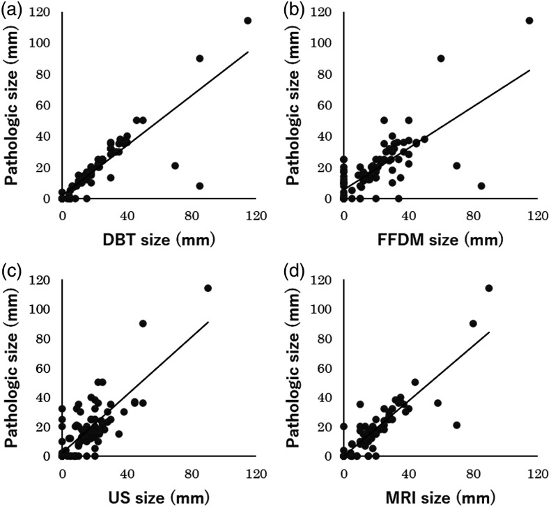
Results: Mean residual tumor size after NST was 19.9 mm for DBT, 18.7 mm for FFDM, 16.0 mm for US, and 18.4 mm for MRI, compared with 17.9 mm on pathology. DBT and MRI correlated better with pathology than that of FFDM and US. The ICC values were 0.85, 0.87, 0.74, and 0.77, respectively. Twenty-five patients (26.3%) achieved pCR after NST. For predicting pCR, area under the receiver operating characteristic (ROC) curve for DBT, FFDM, US, and MRI were 0.79, 0.66, 0.68, and 0.77, respectively.
Conclusion: DBT has good correlation with histopathology for measuring residual tumor size after NST. DBT was comparable to MRI in assessing tumor response after completion of NST.
背景:新辅助全身治疗(NST)的目标是减少肿瘤体积,并为评估治疗反应提供预后指标。数字式乳腺断层合成技术(DBT)被开发出来,由于与全视场数字乳房x线照相术(FFDM)相比,它对乳腺癌检测的灵敏度更高,因此在临床环境中越来越受到关注。目的:评价DBT与FFDM、超声(US)和磁共振成像(MRI)相比在评估乳腺癌患者对NST反应方面的准确性。材料和方法:本回顾性研究纳入95例II-III期乳腺癌患者,这些患者接受了NST并随后进行了手术。NST术后用DBT、FFDM、US、MRI测量残余肿瘤最长直径与病理比较。评估肿瘤大小的一致性和相关性,并评估预测病理完全缓解(pCR)的诊断性能。结果:NST后DBT的平均残余肿瘤大小为19.9 mm, FFDM为18.7 mm, US为16.0 mm, MRI为18.4 mm,病理为17.9 mm。DBT和MRI与病理的相关性优于FFDM和US。ICC值分别为0.85、0.87、0.74、0.77。25例患者(26.3%)在NST后获得pCR。用于预测pCR, DBT、FFDM、US和MRI的受试者工作特征(ROC)曲线下面积分别为0.79、0.66、0.68和0.77。结论:DBT测量NST术后残余肿瘤大小与组织病理学有较好的相关性。在评估NST完成后的肿瘤反应方面,DBT与MRI相当。


 分享
分享

 求助内容:
求助内容: 应助结果提醒方式:
 扫码关注我们
