{"title":"Analysis of purple urine bag syndrome by low vacuum scanning electron microscopy.","authors":"Makoto Abe, Masahito Furuichi, Toshihiko Ishimitsu, Akihiro Tojo","doi":"10.1007/s00795-022-00313-0","DOIUrl":null,"url":null,"abstract":"<p><p>Purple urine bag syndrome (PUBS) is seen in the prolonged indwelling bladder catheters, and the mechanism of its onset was investigated using low vacuum scanning electron microscopy (LVSEM), which enables us to study the 3D structure of urinary sediments and urine bag walls. The urinary sediment and urine bags of 2 cases of PUBS were observed by LVSEM. The urine was brown turbid urine with a pH of 8.5, and magnesium phosphate stones and granules were observed in the urinary sediment together with Gram-positive and Gram-negative bacilli. Bacteria that moved by Brownian motion were observed with a dark-field microscope. LVSEM showed granular crystals around the bacilli, cocci, or mycelium that adhered to the walls of the bag. Granular crystals were dissolved in chloroform and presumed to be a mixture of the bacterial metabolites indigo blue and indirubin red. LVSEM also detected unusual tubular and honeycomb-like graphene in the urinary sediments, which were derived from the inner layer of the silicon elastomer-coated rubber catheter. LVSEM revealed purple crystals produced by bacteria or fungi attached to the urine bag that caused PUBS.</p>","PeriodicalId":18338,"journal":{"name":"Medical Molecular Morphology","volume":"55 2","pages":"123-130"},"PeriodicalIF":1.1000,"publicationDate":"2022-06-01","publicationTypes":"Journal Article","fieldsOfStudy":null,"isOpenAccess":false,"openAccessPdf":"https://www.ncbi.nlm.nih.gov/pmc/articles/PMC9132813/pdf/","citationCount":"2","resultStr":null,"platform":"Semanticscholar","paperid":null,"PeriodicalName":"Medical Molecular Morphology","FirstCategoryId":"3","ListUrlMain":"https://doi.org/10.1007/s00795-022-00313-0","RegionNum":4,"RegionCategory":"医学","ArticlePicture":[],"TitleCN":null,"AbstractTextCN":null,"PMCID":null,"EPubDate":"2022/2/4 0:00:00","PubModel":"Epub","JCR":"Q3","JCRName":"PATHOLOGY","Score":null,"Total":0}
引用次数: 2
Abstract
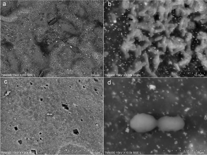
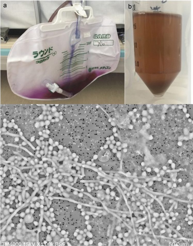
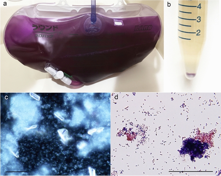
Purple urine bag syndrome (PUBS) is seen in the prolonged indwelling bladder catheters, and the mechanism of its onset was investigated using low vacuum scanning electron microscopy (LVSEM), which enables us to study the 3D structure of urinary sediments and urine bag walls. The urinary sediment and urine bags of 2 cases of PUBS were observed by LVSEM. The urine was brown turbid urine with a pH of 8.5, and magnesium phosphate stones and granules were observed in the urinary sediment together with Gram-positive and Gram-negative bacilli. Bacteria that moved by Brownian motion were observed with a dark-field microscope. LVSEM showed granular crystals around the bacilli, cocci, or mycelium that adhered to the walls of the bag. Granular crystals were dissolved in chloroform and presumed to be a mixture of the bacterial metabolites indigo blue and indirubin red. LVSEM also detected unusual tubular and honeycomb-like graphene in the urinary sediments, which were derived from the inner layer of the silicon elastomer-coated rubber catheter. LVSEM revealed purple crystals produced by bacteria or fungi attached to the urine bag that caused PUBS.
紫色尿袋综合征(Purple urine bag syndrome, PUBS)常见于膀胱导尿管留置时间过长,我们利用低真空扫描电镜(LVSEM)对其发病机制进行了研究,使我们能够研究尿沉积物和尿袋壁的三维结构。用LVSEM对2例酒馆患者的尿沉渣和尿袋进行了观察。尿液呈棕色浑浊,pH为8.5,尿中可见磷酸镁结石和颗粒,革兰氏阳性和革兰氏阴性杆菌。用暗场显微镜观察按布朗运动运动的细菌。LVSEM显示附着在袋壁上的杆菌、球菌或菌丝体周围有颗粒状晶体。颗粒状晶体溶解在氯仿中,推测是细菌代谢物靛蓝和靛红色的混合物。LVSEM还在尿液沉积物中检测到不寻常的管状和蜂窝状石墨烯,这些石墨烯来自于硅弹性体涂层橡胶导管的内层。LVSEM显示,附着在尿袋上的细菌或真菌产生的紫色晶体导致了酒馆。
期刊介绍:
Medical Molecular Morphology is an international forum for researchers in both basic and clinical medicine to present and discuss new research on the structural mechanisms and the processes of health and disease at the molecular level. The structures of molecules, organelles, cells, tissues, and organs determine their normal function. Disease is thus best understood in terms of structural changes in these different levels of biological organization, especially in molecules and molecular interactions as well as the cellular localization of chemical components. Medical Molecular Morphology welcomes articles on basic or clinical research in the fields of cell biology, molecular biology, and medical, veterinary, and dental sciences using techniques for structural research such as electron microscopy, confocal laser scanning microscopy, enzyme histochemistry, immunohistochemistry, radioautography, X-ray microanalysis, and in situ hybridization.
Manuscripts submitted for publication must contain a statement to the effect that all human studies have been reviewed by the appropriate ethics committee and have therefore been performed in accordance with the ethical standards laid down in an appropriate version of the 1964 Declaration of Helsinki. It should also be stated clearly in the text that all persons gave their informed consent prior to their inclusion in the study. Details that might disclose the identity of the subjects under study should be omitted.


 分享
分享
 求助内容:
求助内容: 应助结果提醒方式:
 扫码关注我们
