Investigation of hole-free phase plate performance in transmission electron microscopy under different operation conditions by experiments and simulations
Rebecca Pretzsch, Manuel Dries, Simon Hettler, Martin Spiecker, Martin Obermair, Dagmar Gerthsen
{"title":"Investigation of hole-free phase plate performance in transmission electron microscopy under different operation conditions by experiments and simulations","authors":"Rebecca Pretzsch, Manuel Dries, Simon Hettler, Martin Spiecker, Martin Obermair, Dagmar Gerthsen","doi":"10.1186/s40679-019-0067-z","DOIUrl":null,"url":null,"abstract":"<p>Hole-free phase plates (HFPPs), also known as Volta phase plates, were already demonstrated to be well suited for in-focus transmission electron microscopy imaging of organic objects. However, the underlying physical processes have not been fully understood yet. To further elucidate the imaging properties of HFPPs, phase shift measurements were carried out under different experimental conditions. Both positive and negative phase shifts occur depending on the diameter of the zero-order electron beam and the HFPP film temperature. The analysis of Thon ring patterns of an amorphous carbon test sample reveals that the phase-shifting patch can be significantly larger than the size of the zero-order beam on the HFPP film. An HFPP was used for in-focus phase contrast imaging of carbon nanotube (CNT) bundles under positive and negative phase-shifting conditions. The comparison of experimental and simulated images of CNT bundles gives detailed information on the phase shift profile, which depends on the spatial frequency in the vicinity of the zero-order beam. The shape of the phase shift profile also explains halo-like image artifacts that surround the imaged objects.</p>","PeriodicalId":460,"journal":{"name":"Advanced Structural and Chemical Imaging","volume":"5 1","pages":""},"PeriodicalIF":3.5600,"publicationDate":"2019-10-01","publicationTypes":"Journal Article","fieldsOfStudy":null,"isOpenAccess":false,"openAccessPdf":"https://sci-hub-pdf.com/10.1186/s40679-019-0067-z","citationCount":"7","resultStr":null,"platform":"Semanticscholar","paperid":null,"PeriodicalName":"Advanced Structural and Chemical Imaging","FirstCategoryId":"1085","ListUrlMain":"https://link.springer.com/article/10.1186/s40679-019-0067-z","RegionNum":0,"RegionCategory":null,"ArticlePicture":[],"TitleCN":null,"AbstractTextCN":null,"PMCID":null,"EPubDate":"","PubModel":"","JCR":"Q1","JCRName":"Medicine","Score":null,"Total":0}
引用次数: 7
Abstract
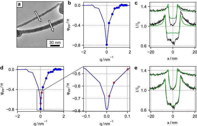
Hole-free phase plates (HFPPs), also known as Volta phase plates, were already demonstrated to be well suited for in-focus transmission electron microscopy imaging of organic objects. However, the underlying physical processes have not been fully understood yet. To further elucidate the imaging properties of HFPPs, phase shift measurements were carried out under different experimental conditions. Both positive and negative phase shifts occur depending on the diameter of the zero-order electron beam and the HFPP film temperature. The analysis of Thon ring patterns of an amorphous carbon test sample reveals that the phase-shifting patch can be significantly larger than the size of the zero-order beam on the HFPP film. An HFPP was used for in-focus phase contrast imaging of carbon nanotube (CNT) bundles under positive and negative phase-shifting conditions. The comparison of experimental and simulated images of CNT bundles gives detailed information on the phase shift profile, which depends on the spatial frequency in the vicinity of the zero-order beam. The shape of the phase shift profile also explains halo-like image artifacts that surround the imaged objects.


 分享
分享
 求助内容:
求助内容: 应助结果提醒方式:
 扫码关注我们
