Imaging of polymer:fullerene bulk-heterojunctions in a scanning electron microscope: methodology aspects and nanomorphology by correlative SEM and STEM
Yonghe Li, Erich Müller, Christian Sprau, Alexander Colsmann, Dagmar Gerthsen
{"title":"Imaging of polymer:fullerene bulk-heterojunctions in a scanning electron microscope: methodology aspects and nanomorphology by correlative SEM and STEM","authors":"Yonghe Li, Erich Müller, Christian Sprau, Alexander Colsmann, Dagmar Gerthsen","doi":"10.1186/s40679-020-00069-4","DOIUrl":null,"url":null,"abstract":"<p>Scanning transmission electron microscopy (STEM) at low energies (≤?30?keV) in a scanning electron microscope is well suited to distinguish weakly scattering materials with similar materials properties and analyze their microstructure. The capabilities of the technique are illustrated in this work to resolve material domains in PTB7:PC<sub>71</sub>BM bulk-heterojunctions, which are commonly implemented for light-harvesting in organic solar cells. Bright-field (BF-) and high-angle annular dark-field (HAADF-) STEM contrast of pure PTB7 and PC<sub>71</sub>BM was first systematically analyzed using a wedge-shaped sample with well-known thickness profile. Monte-Carlo simulations are essential for the assignment of material contrast for materials with only slightly different scattering properties. Different scattering cross-sections were tested in Monte-Carlo simulations with screened Rutherford scattering cross-sections yielding best agreement with the experimental data. The STEM intensity also depends on the local specimen thickness, which can be dealt with by correlative STEM and scanning electron microscopy (SEM) imaging of the same specimen region yielding additional topography information. Correlative STEM/SEM was applied to determine the size of donor (PTB7) and acceptor (PC<sub>71</sub>BM) domains in PTB7:PC<sub>71</sub>BM absorber layers that were deposited from solution with different contents of the processing additive 1,8-diiodooctane (DIO).</p>","PeriodicalId":460,"journal":{"name":"Advanced Structural and Chemical Imaging","volume":"6 1","pages":""},"PeriodicalIF":3.5600,"publicationDate":"2020-03-04","publicationTypes":"Journal Article","fieldsOfStudy":null,"isOpenAccess":false,"openAccessPdf":"https://sci-hub-pdf.com/10.1186/s40679-020-00069-4","citationCount":"3","resultStr":null,"platform":"Semanticscholar","paperid":null,"PeriodicalName":"Advanced Structural and Chemical Imaging","FirstCategoryId":"1085","ListUrlMain":"https://link.springer.com/article/10.1186/s40679-020-00069-4","RegionNum":0,"RegionCategory":null,"ArticlePicture":[],"TitleCN":null,"AbstractTextCN":null,"PMCID":null,"EPubDate":"","PubModel":"","JCR":"Q1","JCRName":"Medicine","Score":null,"Total":0}
引用次数: 3
Abstract
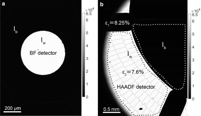
Scanning transmission electron microscopy (STEM) at low energies (≤?30?keV) in a scanning electron microscope is well suited to distinguish weakly scattering materials with similar materials properties and analyze their microstructure. The capabilities of the technique are illustrated in this work to resolve material domains in PTB7:PC71BM bulk-heterojunctions, which are commonly implemented for light-harvesting in organic solar cells. Bright-field (BF-) and high-angle annular dark-field (HAADF-) STEM contrast of pure PTB7 and PC71BM was first systematically analyzed using a wedge-shaped sample with well-known thickness profile. Monte-Carlo simulations are essential for the assignment of material contrast for materials with only slightly different scattering properties. Different scattering cross-sections were tested in Monte-Carlo simulations with screened Rutherford scattering cross-sections yielding best agreement with the experimental data. The STEM intensity also depends on the local specimen thickness, which can be dealt with by correlative STEM and scanning electron microscopy (SEM) imaging of the same specimen region yielding additional topography information. Correlative STEM/SEM was applied to determine the size of donor (PTB7) and acceptor (PC71BM) domains in PTB7:PC71BM absorber layers that were deposited from solution with different contents of the processing additive 1,8-diiodooctane (DIO).


 分享
分享
 求助内容:
求助内容: 应助结果提醒方式:
 扫码关注我们
