{"title":"Removing non-nuclei information from histopathological images: A preprocessing step towards improving nuclei segmentation methods","authors":"Ricardo Moncayo , Anne L. Martel , Eduardo Romero","doi":"10.1016/j.jpi.2023.100315","DOIUrl":null,"url":null,"abstract":"<div><p>Disease interpretation by computer-aided diagnosis systems in digital pathology depends on reliable detection and segmentation of nuclei in hematoxylin and eosin (HE) images. These 2 tasks are challenging since appearance of both cell nuclei and background structures are very variable. This paper presents a method to improve nuclei detection and segmentation in HE images by removing tiles that only contain background information. The method divides each image into smaller patches and uses their projection to the noiselet space to capture different spatial features from non-nuclei background and nuclei structures. The noiselet features are clustered by a <em>K</em>-means algorithm and the resultant partition, defined by the cluster centroids, is herein named the noiselet code-book. A part of an image, a tile, is divided into patches and represented by the histogram of occurrences of the projected patches in the noiselet code-book. Finally, with these histograms, a classifier learns to differentiate between nuclei and non-nuclei tiles. By applying a conventional watershed-marked method to detect and segment nuclei, evaluation consisted in comparing pure watershed method against denoising-plus-watershed in an open database with 8 different types of tissues. The averaged F-score of nuclei detection improved from 0.830 to 0.86 and the dice score after segmentation increased from 0.701 to 0.723.</p></div>","PeriodicalId":37769,"journal":{"name":"Journal of Pathology Informatics","volume":"14 ","pages":"Article 100315"},"PeriodicalIF":0.0000,"publicationDate":"2023-01-01","publicationTypes":"Journal Article","fieldsOfStudy":null,"isOpenAccess":false,"openAccessPdf":"https://www.ncbi.nlm.nih.gov/pmc/articles/PMC10550762/pdf/","citationCount":"0","resultStr":null,"platform":"Semanticscholar","paperid":null,"PeriodicalName":"Journal of Pathology Informatics","FirstCategoryId":"1085","ListUrlMain":"https://www.sciencedirect.com/science/article/pii/S2153353923001293","RegionNum":0,"RegionCategory":null,"ArticlePicture":[],"TitleCN":null,"AbstractTextCN":null,"PMCID":null,"EPubDate":"2023/4/18 0:00:00","PubModel":"Epub","JCR":"Q2","JCRName":"Medicine","Score":null,"Total":0}
引用次数: 0
Abstract
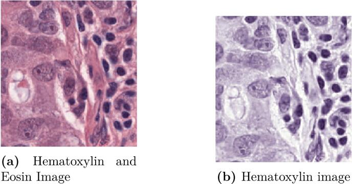
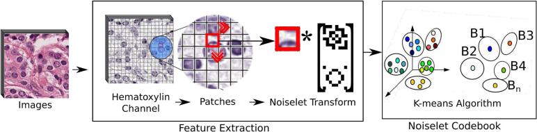
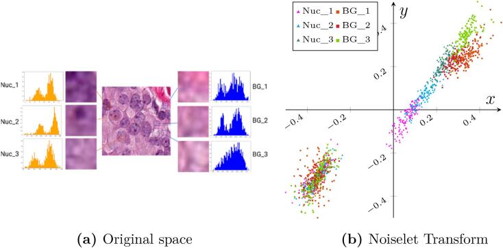
Disease interpretation by computer-aided diagnosis systems in digital pathology depends on reliable detection and segmentation of nuclei in hematoxylin and eosin (HE) images. These 2 tasks are challenging since appearance of both cell nuclei and background structures are very variable. This paper presents a method to improve nuclei detection and segmentation in HE images by removing tiles that only contain background information. The method divides each image into smaller patches and uses their projection to the noiselet space to capture different spatial features from non-nuclei background and nuclei structures. The noiselet features are clustered by a K-means algorithm and the resultant partition, defined by the cluster centroids, is herein named the noiselet code-book. A part of an image, a tile, is divided into patches and represented by the histogram of occurrences of the projected patches in the noiselet code-book. Finally, with these histograms, a classifier learns to differentiate between nuclei and non-nuclei tiles. By applying a conventional watershed-marked method to detect and segment nuclei, evaluation consisted in comparing pure watershed method against denoising-plus-watershed in an open database with 8 different types of tissues. The averaged F-score of nuclei detection improved from 0.830 to 0.86 and the dice score after segmentation increased from 0.701 to 0.723.
期刊介绍:
The Journal of Pathology Informatics (JPI) is an open access peer-reviewed journal dedicated to the advancement of pathology informatics. This is the official journal of the Association for Pathology Informatics (API). The journal aims to publish broadly about pathology informatics and freely disseminate all articles worldwide. This journal is of interest to pathologists, informaticians, academics, researchers, health IT specialists, information officers, IT staff, vendors, and anyone with an interest in informatics. We encourage submissions from anyone with an interest in the field of pathology informatics. We publish all types of papers related to pathology informatics including original research articles, technical notes, reviews, viewpoints, commentaries, editorials, symposia, meeting abstracts, book reviews, and correspondence to the editors. All submissions are subject to rigorous peer review by the well-regarded editorial board and by expert referees in appropriate specialties.


 分享
分享
 求助内容:
求助内容: 应助结果提醒方式:
 扫码关注我们
