W. A. Moeglein, R. Griswold, B. L. Mehdi, N. D. Browning, J. Teuton
{"title":"Applying shot boundary detection for automated crystal growth analysis during in situ transmission electron microscope experiments","authors":"W. A. Moeglein, R. Griswold, B. L. Mehdi, N. D. Browning, J. Teuton","doi":"10.1186/s40679-016-0034-x","DOIUrl":null,"url":null,"abstract":"<p>In situ scanning transmission electron microscopy is being developed for numerous applications in the study of nucleation and growth under electrochemical driving forces. For this type of experiment, one of the key parameters is to identify when nucleation initiates. Typically, the process of identifying the moment that crystals begin to form is a manual process requiring the user to perform an observation and respond accordingly (adjust focus, magnification, translate the stage, etc.). However, as the speed of the cameras being used to perform these observations increases, the ability of a user to “catch” the important initial stage of nucleation decreases (there is more information that is available in the first few milliseconds of the process). Here, we show that video shot boundary detection can automatically detect frames where a change in the image occurs. We show that this method can be applied to quickly and accurately identify points of change during crystal growth. This technique allows for automated segmentation of a digital stream for further analysis and the assignment of arbitrary time stamps for the initiation of processes that are independent of the user’s ability to observe and react.</p>","PeriodicalId":460,"journal":{"name":"Advanced Structural and Chemical Imaging","volume":"3 1","pages":""},"PeriodicalIF":3.5600,"publicationDate":"2017-01-03","publicationTypes":"Journal Article","fieldsOfStudy":null,"isOpenAccess":false,"openAccessPdf":"https://sci-hub-pdf.com/10.1186/s40679-016-0034-x","citationCount":"7","resultStr":null,"platform":"Semanticscholar","paperid":null,"PeriodicalName":"Advanced Structural and Chemical Imaging","FirstCategoryId":"1085","ListUrlMain":"https://link.springer.com/article/10.1186/s40679-016-0034-x","RegionNum":0,"RegionCategory":null,"ArticlePicture":[],"TitleCN":null,"AbstractTextCN":null,"PMCID":null,"EPubDate":"","PubModel":"","JCR":"Q1","JCRName":"Medicine","Score":null,"Total":0}
引用次数: 7
Abstract
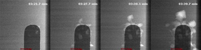
In situ scanning transmission electron microscopy is being developed for numerous applications in the study of nucleation and growth under electrochemical driving forces. For this type of experiment, one of the key parameters is to identify when nucleation initiates. Typically, the process of identifying the moment that crystals begin to form is a manual process requiring the user to perform an observation and respond accordingly (adjust focus, magnification, translate the stage, etc.). However, as the speed of the cameras being used to perform these observations increases, the ability of a user to “catch” the important initial stage of nucleation decreases (there is more information that is available in the first few milliseconds of the process). Here, we show that video shot boundary detection can automatically detect frames where a change in the image occurs. We show that this method can be applied to quickly and accurately identify points of change during crystal growth. This technique allows for automated segmentation of a digital stream for further analysis and the assignment of arbitrary time stamps for the initiation of processes that are independent of the user’s ability to observe and react.


 分享
分享
 求助内容:
求助内容: 应助结果提醒方式:
 扫码关注我们
