Rhia M Martin, Samantha Diaz, Martin Poncelet, Benoit Driesschaert, Eugene Barth, Mrignayani Kotecha, Boris Epel, Gareth R Eaton, Joshua R Biller
{"title":"Toward a Nanoencapsulated EPR Imaging Agent for Clinical Use.","authors":"Rhia M Martin, Samantha Diaz, Martin Poncelet, Benoit Driesschaert, Eugene Barth, Mrignayani Kotecha, Boris Epel, Gareth R Eaton, Joshua R Biller","doi":"10.1007/s11307-023-01863-0","DOIUrl":null,"url":null,"abstract":"<p><strong>Purpose: </strong>Progress toward developing a novel radiocontrast agent for determining pO<sub>2</sub> in tumors in a clinical setting is described. The imaging agent is designed for use with electron paramagnetic resonance imaging (EPRI), in which the collision of a paramagnetic probe molecule with molecular oxygen causes a spectroscopic change which can be calibrated to give the real oxygen concentration in the tumor tissue.</p><p><strong>Procedures: </strong>The imaging agent is based on a nanoscaffold of aluminum hydroxide (boehmite) with sizes from 100 to 200 nm, paramagnetic probe molecule, and encapsulation with a gas permeable, thin (10-20 nm) polymer layer to separate the imaging agent and body environment while still allowing O<sub>2</sub> to interact with the paramagnetic probe. A specially designed deuterated Finland trityl (dFT) is covalently attached on the surface of the nanoparticle through 1,3-dipolar addition of the alkyne on the dFT with an azide on the surface of the nanoscaffold. This click-chemistry reaction affords 100% efficiency of the trityl attachment as followed by the complete disappearance of the azide peak in the infrared spectrum. The fully encapsulated, dFT-functionalized nanoparticle is referred to as RADI-Sense.</p><p><strong>Results: </strong>Side-by-side in vivo imaging comparisons made in a mouse model made between RADI-Sense and free paramagnetic probe (OX-071) showed oxygen sensitivity is retained and RADI-Sense can create 3D pO<sub>2</sub> maps of solid tumors CONCLUSIONS: A novel encapsulated nanoparticle EPR imaging agent has been described which could be used in the future to bring EPR imaging for guidance of radiotherapy into clinical reality.</p>","PeriodicalId":18760,"journal":{"name":"Molecular Imaging and Biology","volume":" ","pages":"525-541"},"PeriodicalIF":2.5000,"publicationDate":"2024-06-01","publicationTypes":"Journal Article","fieldsOfStudy":null,"isOpenAccess":false,"openAccessPdf":"https://www.ncbi.nlm.nih.gov/pmc/articles/PMC11035482/pdf/","citationCount":"0","resultStr":null,"platform":"Semanticscholar","paperid":null,"PeriodicalName":"Molecular Imaging and Biology","FirstCategoryId":"3","ListUrlMain":"https://doi.org/10.1007/s11307-023-01863-0","RegionNum":4,"RegionCategory":"医学","ArticlePicture":[],"TitleCN":null,"AbstractTextCN":null,"PMCID":null,"EPubDate":"2023/10/23 0:00:00","PubModel":"Epub","JCR":"Q2","JCRName":"RADIOLOGY, NUCLEAR MEDICINE & MEDICAL IMAGING","Score":null,"Total":0}
引用次数: 0
Abstract
Purpose: Progress toward developing a novel radiocontrast agent for determining pO2 in tumors in a clinical setting is described. The imaging agent is designed for use with electron paramagnetic resonance imaging (EPRI), in which the collision of a paramagnetic probe molecule with molecular oxygen causes a spectroscopic change which can be calibrated to give the real oxygen concentration in the tumor tissue.
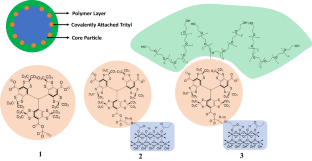
Procedures: The imaging agent is based on a nanoscaffold of aluminum hydroxide (boehmite) with sizes from 100 to 200 nm, paramagnetic probe molecule, and encapsulation with a gas permeable, thin (10-20 nm) polymer layer to separate the imaging agent and body environment while still allowing O2 to interact with the paramagnetic probe. A specially designed deuterated Finland trityl (dFT) is covalently attached on the surface of the nanoparticle through 1,3-dipolar addition of the alkyne on the dFT with an azide on the surface of the nanoscaffold. This click-chemistry reaction affords 100% efficiency of the trityl attachment as followed by the complete disappearance of the azide peak in the infrared spectrum. The fully encapsulated, dFT-functionalized nanoparticle is referred to as RADI-Sense.
Results: Side-by-side in vivo imaging comparisons made in a mouse model made between RADI-Sense and free paramagnetic probe (OX-071) showed oxygen sensitivity is retained and RADI-Sense can create 3D pO2 maps of solid tumors CONCLUSIONS: A novel encapsulated nanoparticle EPR imaging agent has been described which could be used in the future to bring EPR imaging for guidance of radiotherapy into clinical reality.
期刊介绍:
Molecular Imaging and Biology (MIB) invites original contributions (research articles, review articles, commentaries, etc.) on the utilization of molecular imaging (i.e., nuclear imaging, optical imaging, autoradiography and pathology, MRI, MPI, ultrasound imaging, radiomics/genomics etc.) to investigate questions related to biology and health. The objective of MIB is to provide a forum to the discovery of molecular mechanisms of disease through the use of imaging techniques. We aim to investigate the biological nature of disease in patients and establish new molecular imaging diagnostic and therapy procedures.
Some areas that are covered are:
Preclinical and clinical imaging of macromolecular targets (e.g., genes, receptors, enzymes) involved in significant biological processes.
The design, characterization, and study of new molecular imaging probes and contrast agents for the functional interrogation of macromolecular targets.
Development and evaluation of imaging systems including instrumentation, image reconstruction algorithms, image analysis, and display.
Development of molecular assay approaches leading to quantification of the biological information obtained in molecular imaging.
Study of in vivo animal models of disease for the development of new molecular diagnostics and therapeutics.
Extension of in vitro and in vivo discoveries using disease models, into well designed clinical research investigations.
Clinical molecular imaging involving clinical investigations, clinical trials and medical management or cost-effectiveness studies.


 分享
分享

 求助内容:
求助内容: 应助结果提醒方式:
 扫码关注我们
