Hanan B Elajaili, Nathan M Dee, Sergey I Dikalov, Joseph P Y Kao, Eva S Nozik
{"title":"Use of Electron Paramagnetic Resonance (EPR) to Evaluate Redox Status in a Preclinical Model of Acute Lung Injury.","authors":"Hanan B Elajaili, Nathan M Dee, Sergey I Dikalov, Joseph P Y Kao, Eva S Nozik","doi":"10.1007/s11307-023-01826-5","DOIUrl":null,"url":null,"abstract":"<p><strong>Purpose: </strong>Patients with hyper- vs. hypo-inflammatory subphenotypes of acute respiratory distress syndrome (ARDS) exhibit different clinical outcomes. Inflammation increases the production of reactive oxygen species (ROS) and increased ROS contributes to the severity of illness. Our long-term goal is to develop electron paramagnetic resonance (EPR) imaging of lungs in vivo to precisely measure superoxide production in ARDS in real time. As a first step, this requires the development of in vivo EPR methods for quantifying superoxide generation in the lung during injury, and testing if such superoxide measurements can differentiate between susceptible and protected mouse strains.</p><p><strong>Procedures: </strong>In WT mice, mice lacking total body extracellular superoxide dismutase (EC-SOD) (KO), or mice overexpressing lung EC-SOD (Tg), lung injury was induced with intraperitoneal (IP) lipopolysaccharide (LPS) (10 mg/kg). At 24 h after LPS treatment, mice were injected with the cyclic hydroxylamines 1-hydroxy-3-carboxy-2,2,5,5-tetramethylpyrrolidine hydrochloride (CPH) or 4-acetoxymethoxycarbonyl-1-hydroxy-2,2,5,5-tetramethylpyrrolidine-3-carboxylic acid (DCP-AM-H) probes to detect, respectively, cellular and mitochondrial ROS - specifically superoxide. Several probe delivery strategies were tested. Lung tissue was collected up to one hour after probe administration and assayed by EPR.</p><p><strong>Results: </strong>As measured by X-band EPR, cellular and mitochondrial superoxide increased in the lungs of LPS-treated mice compared to control. Lung cellular superoxide was increased in EC-SOD KO mice and decreased in EC-SOD Tg mice compared to WT. We also validated an intratracheal (IT) delivery method, which enhanced the lung signal for both spin probes compared to IP administration.</p><p><strong>Conclusions: </strong>We have developed protocols for delivering EPR spin probes in vivo, allowing detection of cellular and mitochondrial superoxide in lung injury by EPR. Superoxide measurements by EPR could differentiate mice with and without lung injury, as well as mouse strains with different disease susceptibilities. We expect these protocols to capture real-time superoxide production and enable evaluation of lung EPR imaging as a potential clinical tool for subphenotyping ARDS patients based on redox status.</p>","PeriodicalId":18760,"journal":{"name":"Molecular Imaging and Biology","volume":" ","pages":"495-502"},"PeriodicalIF":2.5000,"publicationDate":"2024-06-01","publicationTypes":"Journal Article","fieldsOfStudy":null,"isOpenAccess":false,"openAccessPdf":"https://www.ncbi.nlm.nih.gov/pmc/articles/PMC10188229/pdf/","citationCount":"0","resultStr":null,"platform":"Semanticscholar","paperid":null,"PeriodicalName":"Molecular Imaging and Biology","FirstCategoryId":"3","ListUrlMain":"https://doi.org/10.1007/s11307-023-01826-5","RegionNum":4,"RegionCategory":"医学","ArticlePicture":[],"TitleCN":null,"AbstractTextCN":null,"PMCID":null,"EPubDate":"2023/5/16 0:00:00","PubModel":"Epub","JCR":"Q2","JCRName":"RADIOLOGY, NUCLEAR MEDICINE & MEDICAL IMAGING","Score":null,"Total":0}
引用次数: 0
Abstract
Purpose: Patients with hyper- vs. hypo-inflammatory subphenotypes of acute respiratory distress syndrome (ARDS) exhibit different clinical outcomes. Inflammation increases the production of reactive oxygen species (ROS) and increased ROS contributes to the severity of illness. Our long-term goal is to develop electron paramagnetic resonance (EPR) imaging of lungs in vivo to precisely measure superoxide production in ARDS in real time. As a first step, this requires the development of in vivo EPR methods for quantifying superoxide generation in the lung during injury, and testing if such superoxide measurements can differentiate between susceptible and protected mouse strains.
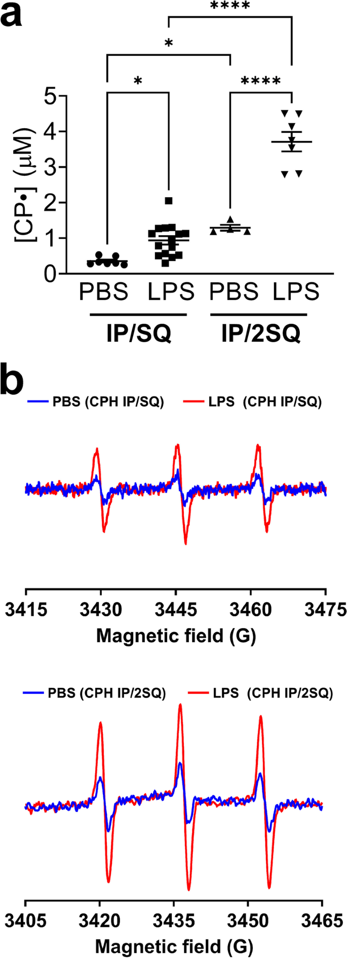
Procedures: In WT mice, mice lacking total body extracellular superoxide dismutase (EC-SOD) (KO), or mice overexpressing lung EC-SOD (Tg), lung injury was induced with intraperitoneal (IP) lipopolysaccharide (LPS) (10 mg/kg). At 24 h after LPS treatment, mice were injected with the cyclic hydroxylamines 1-hydroxy-3-carboxy-2,2,5,5-tetramethylpyrrolidine hydrochloride (CPH) or 4-acetoxymethoxycarbonyl-1-hydroxy-2,2,5,5-tetramethylpyrrolidine-3-carboxylic acid (DCP-AM-H) probes to detect, respectively, cellular and mitochondrial ROS - specifically superoxide. Several probe delivery strategies were tested. Lung tissue was collected up to one hour after probe administration and assayed by EPR.
Results: As measured by X-band EPR, cellular and mitochondrial superoxide increased in the lungs of LPS-treated mice compared to control. Lung cellular superoxide was increased in EC-SOD KO mice and decreased in EC-SOD Tg mice compared to WT. We also validated an intratracheal (IT) delivery method, which enhanced the lung signal for both spin probes compared to IP administration.
Conclusions: We have developed protocols for delivering EPR spin probes in vivo, allowing detection of cellular and mitochondrial superoxide in lung injury by EPR. Superoxide measurements by EPR could differentiate mice with and without lung injury, as well as mouse strains with different disease susceptibilities. We expect these protocols to capture real-time superoxide production and enable evaluation of lung EPR imaging as a potential clinical tool for subphenotyping ARDS patients based on redox status.
期刊介绍:
Molecular Imaging and Biology (MIB) invites original contributions (research articles, review articles, commentaries, etc.) on the utilization of molecular imaging (i.e., nuclear imaging, optical imaging, autoradiography and pathology, MRI, MPI, ultrasound imaging, radiomics/genomics etc.) to investigate questions related to biology and health. The objective of MIB is to provide a forum to the discovery of molecular mechanisms of disease through the use of imaging techniques. We aim to investigate the biological nature of disease in patients and establish new molecular imaging diagnostic and therapy procedures.
Some areas that are covered are:
Preclinical and clinical imaging of macromolecular targets (e.g., genes, receptors, enzymes) involved in significant biological processes.
The design, characterization, and study of new molecular imaging probes and contrast agents for the functional interrogation of macromolecular targets.
Development and evaluation of imaging systems including instrumentation, image reconstruction algorithms, image analysis, and display.
Development of molecular assay approaches leading to quantification of the biological information obtained in molecular imaging.
Study of in vivo animal models of disease for the development of new molecular diagnostics and therapeutics.
Extension of in vitro and in vivo discoveries using disease models, into well designed clinical research investigations.
Clinical molecular imaging involving clinical investigations, clinical trials and medical management or cost-effectiveness studies.


 分享
分享

 求助内容:
求助内容: 应助结果提醒方式:
 扫码关注我们
