Maria Piagkou, Aliki Fiska, George Tsakotos, George Triantafyllou, Constantinus Politis, Christos Koutserimpas, Janusz Skrzat, Lukasz Olewnik, Nicole Zielinska, Athina Tousia, Michael Kostares, Trifon Totlis, Anastasia Triantafyllou, Katerina Al Nasraoui, Vasilios Karampelias, Christos Tsiouris, Konstantinos Natsis
{"title":"A morphological study on the sphenoid bone ligaments' ossification pattern.","authors":"Maria Piagkou, Aliki Fiska, George Tsakotos, George Triantafyllou, Constantinus Politis, Christos Koutserimpas, Janusz Skrzat, Lukasz Olewnik, Nicole Zielinska, Athina Tousia, Michael Kostares, Trifon Totlis, Anastasia Triantafyllou, Katerina Al Nasraoui, Vasilios Karampelias, Christos Tsiouris, Konstantinos Natsis","doi":"10.1007/s00276-023-03226-4","DOIUrl":null,"url":null,"abstract":"<p><strong>Purpose: </strong>The sphenoid bone (SB) extracranial ligaments (ECRLs) are the pterygoalar and pterygospinous ligaments (PTAL and PTSL) that are located at the SB lateral pterygoid plate, and inferior to the foramen ovale (FO). Their ossification may affect the mandibular nerve's distribution. The intracranial ligaments' (ICRLs) ossification (the caroticoclinoid ligament-CCLL, the anterior and posterior interclinoid ligaments-AICLL and PICLL) may impede the approaches to the sella. This study highlights the incidence of the ossified ECRLs and ICRLs location, their type (partial, or complete), considering laterality, gender, age, and ligaments' simultaneous presence.</p><p><strong>Methods: </strong>The sample consisted of 156 Greek adult dried skulls of both genders and variable age.</p><p><strong>Results: </strong>Ossified ligaments were identified in 57.05%, predominantly extracranially (42.31%, P = 0.003). ECRLs were predominantly identified unilaterally (30.13%, P < 0.001). The majority of the ossified ICRLs were predominantly identified in male skulls (31.1%, P = 0.048) and the majority of the ECRLs (52.8%, P = 0.028) were predominantly identified at the age of 60 years and above. The PTAL was the most ossified (32.69%), followed by the CCLL (24.36%), the PTSL (16.03%), the PICLL (6.41%), and the AICLL (4.49%).</p><p><strong>Conclusions: </strong>Detailed knowledge of the SB morphology and ligaments' ossification extent is essential to improve the technique of the FO percutaneous approach, and sellar approaches, to minimize complications.</p>","PeriodicalId":49296,"journal":{"name":"Surgical and Radiologic Anatomy","volume":" ","pages":"1405-1417"},"PeriodicalIF":1.2000,"publicationDate":"2023-11-01","publicationTypes":"Journal Article","fieldsOfStudy":null,"isOpenAccess":false,"openAccessPdf":"https://www.ncbi.nlm.nih.gov/pmc/articles/PMC10587028/pdf/","citationCount":"0","resultStr":null,"platform":"Semanticscholar","paperid":null,"PeriodicalName":"Surgical and Radiologic Anatomy","FirstCategoryId":"3","ListUrlMain":"https://doi.org/10.1007/s00276-023-03226-4","RegionNum":4,"RegionCategory":"医学","ArticlePicture":[],"TitleCN":null,"AbstractTextCN":null,"PMCID":null,"EPubDate":"2023/8/7 0:00:00","PubModel":"Epub","JCR":"Q3","JCRName":"ANATOMY & MORPHOLOGY","Score":null,"Total":0}
引用次数: 0
Abstract
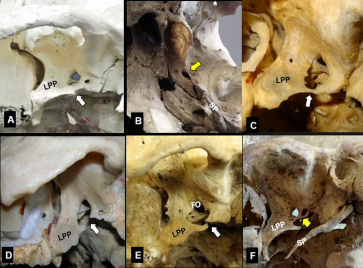
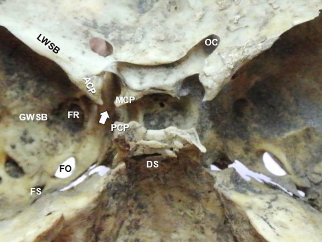
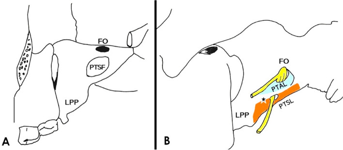
Purpose: The sphenoid bone (SB) extracranial ligaments (ECRLs) are the pterygoalar and pterygospinous ligaments (PTAL and PTSL) that are located at the SB lateral pterygoid plate, and inferior to the foramen ovale (FO). Their ossification may affect the mandibular nerve's distribution. The intracranial ligaments' (ICRLs) ossification (the caroticoclinoid ligament-CCLL, the anterior and posterior interclinoid ligaments-AICLL and PICLL) may impede the approaches to the sella. This study highlights the incidence of the ossified ECRLs and ICRLs location, their type (partial, or complete), considering laterality, gender, age, and ligaments' simultaneous presence.
Methods: The sample consisted of 156 Greek adult dried skulls of both genders and variable age.
Results: Ossified ligaments were identified in 57.05%, predominantly extracranially (42.31%, P = 0.003). ECRLs were predominantly identified unilaterally (30.13%, P < 0.001). The majority of the ossified ICRLs were predominantly identified in male skulls (31.1%, P = 0.048) and the majority of the ECRLs (52.8%, P = 0.028) were predominantly identified at the age of 60 years and above. The PTAL was the most ossified (32.69%), followed by the CCLL (24.36%), the PTSL (16.03%), the PICLL (6.41%), and the AICLL (4.49%).
Conclusions: Detailed knowledge of the SB morphology and ligaments' ossification extent is essential to improve the technique of the FO percutaneous approach, and sellar approaches, to minimize complications.
期刊介绍:
Anatomy is a morphological science which cannot fail to interest the clinician. The practical application of anatomical research to clinical problems necessitates special adaptation and selectivity in choosing from numerous international works. Although there is a tendency to believe that meaningful advances in anatomy are unlikely, constant revision is necessary. Surgical and Radiologic Anatomy, the first international journal of Clinical anatomy has been created in this spirit.
Its goal is to serve clinicians, regardless of speciality-physicians, surgeons, radiologists or other specialists-as an indispensable aid with which they can improve their knowledge of anatomy. Each issue includes: Original papers, review articles, articles on the anatomical bases of medical, surgical and radiological techniques, articles of normal radiologic anatomy, brief reviews of anatomical publications of clinical interest.
Particular attention is given to high quality illustrations, which are indispensable for a better understanding of anatomical problems.
Surgical and Radiologic Anatomy is a journal written by anatomists for clinicians with a special interest in anatomy.


 分享
分享
 求助内容:
求助内容: 应助结果提醒方式:
 扫码关注我们
