{"title":"在3D-FLAIR中使用t2对比增强技术检测内淋巴水肿的缺陷。","authors":"Yutaka Kato, Shinji Naganawa, Toshiaki Taoka, Tadao Yoshida, Michihiko Sone","doi":"10.2463/mrms.mp.2022-0017","DOIUrl":null,"url":null,"abstract":"Purpose To determine whether T2-contrast enhancement techniques can be used to diagnose endolymphatic hydrops, we compared fluid signal artifacts with and without T2-contrast enhancement techniques in 3D fluid-attenuated inversion recovery (3D-FLAIR). Methods We prepared a custom-made phantom consisting of eight tubes half-filled with saline. Images were obtained using four 3D-FLAIR: without T2-contrast enhancement (Normal), with non-selective T2-inversion recovery (T2-IR), and two with non-selective T2 preparation IR (T2-prep). Scans were performed with and without rice covering the phantom to simulate minimal and severe B0-inhomogeneity conditions. The average signal intensity (SI) values of eight saline tubes were compared between the four sequences and between each other. Comparisons were performed for all measurement slices and the central 10 slices. The images using T2-contrast enhancement technique were obtained from a volunteer and a patient suspected of Meniere’s disease. Results The Normal sequence SI for all slices was significantly lower than that for the other sequences, with smaller standard deviation (SD) and no outliers. Several outliers were detected in the other sequences. The SDs and outliers were larger without rice than with rice. When the central 10 slices with rice, the T2-IR had a significantly higher SI with more outliers compared with the Normal sequence. The T2-prep had no outliers and SIs that were comparable to those of the Normal sequence. However, without rice, the T2-IR and T2-prep sequences had significantly higher SIs with outliers and larger SDs compared to the Normal sequence. In the corresponding images, the Normal sequence achieved excellent fluid suppression, whereas the T2-IR and T2-prep sequences showed high-signal artifacts. Imperfect fluid suppressions were observed in the volunteer image and the endolymphatic hydrops on the post-gadolinium image differed in size and shape in the non-injected T2-IR in the patient image. Conclusion T2-contrast enhancement techniques should be used with caution in 3D-FLAIR for diagnosing endolymphatic hydrops.","PeriodicalId":18119,"journal":{"name":"Magnetic Resonance in Medical Sciences","volume":"22 3","pages":"335-344"},"PeriodicalIF":3.2000,"publicationDate":"2023-07-01","publicationTypes":"Journal Article","fieldsOfStudy":null,"isOpenAccess":false,"openAccessPdf":"https://ftp.ncbi.nlm.nih.gov/pub/pmc/oa_pdf/e3/5b/mrms-22-335.PMC10449551.pdf","citationCount":"2","resultStr":"{\"title\":\"Pitfalls of Using T2-contrast Enhancement Techniques in 3D-FLAIR to Detect Endolymphatic Hydrops.\",\"authors\":\"Yutaka Kato, Shinji Naganawa, Toshiaki Taoka, Tadao Yoshida, Michihiko Sone\",\"doi\":\"10.2463/mrms.mp.2022-0017\",\"DOIUrl\":null,\"url\":null,\"abstract\":\"Purpose To determine whether T2-contrast enhancement techniques can be used to diagnose endolymphatic hydrops, we compared fluid signal artifacts with and without T2-contrast enhancement techniques in 3D fluid-attenuated inversion recovery (3D-FLAIR). Methods We prepared a custom-made phantom consisting of eight tubes half-filled with saline. Images were obtained using four 3D-FLAIR: without T2-contrast enhancement (Normal), with non-selective T2-inversion recovery (T2-IR), and two with non-selective T2 preparation IR (T2-prep). Scans were performed with and without rice covering the phantom to simulate minimal and severe B0-inhomogeneity conditions. The average signal intensity (SI) values of eight saline tubes were compared between the four sequences and between each other. Comparisons were performed for all measurement slices and the central 10 slices. The images using T2-contrast enhancement technique were obtained from a volunteer and a patient suspected of Meniere’s disease. Results The Normal sequence SI for all slices was significantly lower than that for the other sequences, with smaller standard deviation (SD) and no outliers. Several outliers were detected in the other sequences. The SDs and outliers were larger without rice than with rice. When the central 10 slices with rice, the T2-IR had a significantly higher SI with more outliers compared with the Normal sequence. The T2-prep had no outliers and SIs that were comparable to those of the Normal sequence. However, without rice, the T2-IR and T2-prep sequences had significantly higher SIs with outliers and larger SDs compared to the Normal sequence. In the corresponding images, the Normal sequence achieved excellent fluid suppression, whereas the T2-IR and T2-prep sequences showed high-signal artifacts. Imperfect fluid suppressions were observed in the volunteer image and the endolymphatic hydrops on the post-gadolinium image differed in size and shape in the non-injected T2-IR in the patient image. Conclusion T2-contrast enhancement techniques should be used with caution in 3D-FLAIR for diagnosing endolymphatic hydrops.\",\"PeriodicalId\":18119,\"journal\":{\"name\":\"Magnetic Resonance in Medical Sciences\",\"volume\":\"22 3\",\"pages\":\"335-344\"},\"PeriodicalIF\":3.2000,\"publicationDate\":\"2023-07-01\",\"publicationTypes\":\"Journal Article\",\"fieldsOfStudy\":null,\"isOpenAccess\":false,\"openAccessPdf\":\"https://ftp.ncbi.nlm.nih.gov/pub/pmc/oa_pdf/e3/5b/mrms-22-335.PMC10449551.pdf\",\"citationCount\":\"2\",\"resultStr\":null,\"platform\":\"Semanticscholar\",\"paperid\":null,\"PeriodicalName\":\"Magnetic Resonance in Medical Sciences\",\"FirstCategoryId\":\"3\",\"ListUrlMain\":\"https://doi.org/10.2463/mrms.mp.2022-0017\",\"RegionNum\":3,\"RegionCategory\":\"医学\",\"ArticlePicture\":[],\"TitleCN\":null,\"AbstractTextCN\":null,\"PMCID\":null,\"EPubDate\":\"\",\"PubModel\":\"\",\"JCR\":\"Q2\",\"JCRName\":\"RADIOLOGY, NUCLEAR MEDICINE & MEDICAL IMAGING\",\"Score\":null,\"Total\":0}","platform":"Semanticscholar","paperid":null,"PeriodicalName":"Magnetic Resonance in Medical Sciences","FirstCategoryId":"3","ListUrlMain":"https://doi.org/10.2463/mrms.mp.2022-0017","RegionNum":3,"RegionCategory":"医学","ArticlePicture":[],"TitleCN":null,"AbstractTextCN":null,"PMCID":null,"EPubDate":"","PubModel":"","JCR":"Q2","JCRName":"RADIOLOGY, NUCLEAR MEDICINE & MEDICAL IMAGING","Score":null,"Total":0}
Pitfalls of Using T2-contrast Enhancement Techniques in 3D-FLAIR to Detect Endolymphatic Hydrops.
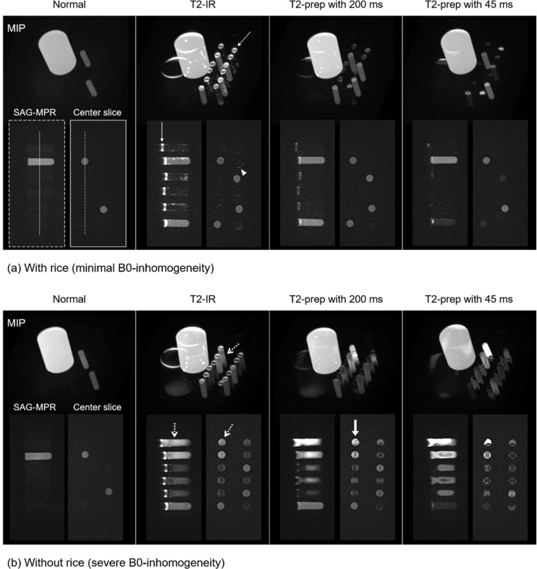
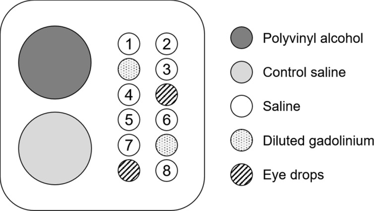
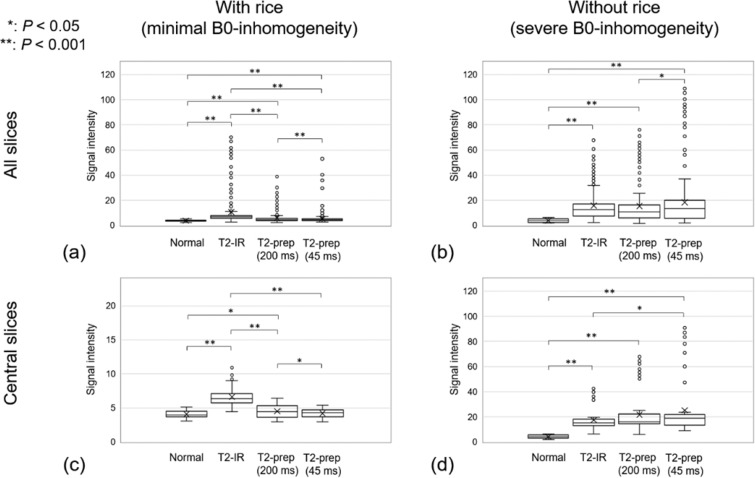
Purpose To determine whether T2-contrast enhancement techniques can be used to diagnose endolymphatic hydrops, we compared fluid signal artifacts with and without T2-contrast enhancement techniques in 3D fluid-attenuated inversion recovery (3D-FLAIR). Methods We prepared a custom-made phantom consisting of eight tubes half-filled with saline. Images were obtained using four 3D-FLAIR: without T2-contrast enhancement (Normal), with non-selective T2-inversion recovery (T2-IR), and two with non-selective T2 preparation IR (T2-prep). Scans were performed with and without rice covering the phantom to simulate minimal and severe B0-inhomogeneity conditions. The average signal intensity (SI) values of eight saline tubes were compared between the four sequences and between each other. Comparisons were performed for all measurement slices and the central 10 slices. The images using T2-contrast enhancement technique were obtained from a volunteer and a patient suspected of Meniere’s disease. Results The Normal sequence SI for all slices was significantly lower than that for the other sequences, with smaller standard deviation (SD) and no outliers. Several outliers were detected in the other sequences. The SDs and outliers were larger without rice than with rice. When the central 10 slices with rice, the T2-IR had a significantly higher SI with more outliers compared with the Normal sequence. The T2-prep had no outliers and SIs that were comparable to those of the Normal sequence. However, without rice, the T2-IR and T2-prep sequences had significantly higher SIs with outliers and larger SDs compared to the Normal sequence. In the corresponding images, the Normal sequence achieved excellent fluid suppression, whereas the T2-IR and T2-prep sequences showed high-signal artifacts. Imperfect fluid suppressions were observed in the volunteer image and the endolymphatic hydrops on the post-gadolinium image differed in size and shape in the non-injected T2-IR in the patient image. Conclusion T2-contrast enhancement techniques should be used with caution in 3D-FLAIR for diagnosing endolymphatic hydrops.
期刊介绍:
Magnetic Resonance in Medical Sciences (MRMS or Magn
Reson Med Sci) is an international journal pursuing the
publication of original articles contributing to the progress
of magnetic resonance in the field of biomedical sciences
including technical developments and clinical applications.
MRMS is an official journal of the Japanese Society for
Magnetic Resonance in Medicine (JSMRM).


 分享
分享
 求助内容:
求助内容: 应助结果提醒方式:
 扫码关注我们
