Cristina Martinez-Fernandez de la Camara, Tina Storm, Ahmed Salman, Thomas Burgoyne, Martin Qvist Rasmussen, Harry O Orlans, Angela J Russell, Stephen G Davies, Alun R Barnard, Robert E MacLaren
{"title":"视网膜胶质细胞中细胞周期调节因子 p16INK4a 的发育表达:未成熟眼星形胶质细胞的新标记?","authors":"Cristina Martinez-Fernandez de la Camara, Tina Storm, Ahmed Salman, Thomas Burgoyne, Martin Qvist Rasmussen, Harry O Orlans, Angela J Russell, Stephen G Davies, Alun R Barnard, Robert E MacLaren","doi":"10.1369/00221554231184286","DOIUrl":null,"url":null,"abstract":"<p><p>Retinal astrocytes are vital for neuronal homeostasis in the retina. Together with Müller glia, they provide retinal cells with neurotrophic factors, antioxidative support, and defense mechanisms such as the formation of the blood-retinal barrier. Substantial heterogeneity of astrocyte morphology and function represents a challenge for identification of distinct subtypes which may be potential targets for therapeutic purposes. Hence, identification of novel markers of astrocyte subpopulations is highly relevant to better understand the molecular mechanisms involved in retinal development, homeostasis, and pathology. In this study, we observed that the cell cycle regulator, p16<sup>INK4a</sup>, is expressed in immature astrocytes in the mouse retina. Immunohistochemical analysis showed p16<sup>INK4a</sup> expression in the optic nerve of wild-type mice from 3 days to 3 months of age and in the nerve fiber layer of the adult mouse retina. Colocalization of p16<sup>INK4a</sup> expression and glial fibrillary acidic protein (immature/mature astrocyte marker) tends to decrease with age. However, colocalization of p16<sup>INK4a</sup> expression and vimentin (immature astrocyte marker) remains high in the optic nerve from the early postnatal period to adulthood. The observations from this study provide a valuable tool for further investigations of ocular astrocytes in the developing retina as well as in degenerative retinopathies.</p>","PeriodicalId":16079,"journal":{"name":"Journal of Histochemistry & Cytochemistry","volume":"71 6","pages":"301-320"},"PeriodicalIF":1.5000,"publicationDate":"2023-06-01","publicationTypes":"Journal Article","fieldsOfStudy":null,"isOpenAccess":false,"openAccessPdf":"https://ftp.ncbi.nlm.nih.gov/pub/pmc/oa_pdf/0a/be/10.1369_00221554231184286.PMC10315990.pdf","citationCount":"0","resultStr":"{\"title\":\"Developmental Expression of the Cell Cycle Regulator p16<sup>INK4a</sup> in Retinal Glial Cells: A Novel Marker for Immature Ocular Astrocytes?\",\"authors\":\"Cristina Martinez-Fernandez de la Camara, Tina Storm, Ahmed Salman, Thomas Burgoyne, Martin Qvist Rasmussen, Harry O Orlans, Angela J Russell, Stephen G Davies, Alun R Barnard, Robert E MacLaren\",\"doi\":\"10.1369/00221554231184286\",\"DOIUrl\":null,\"url\":null,\"abstract\":\"<p><p>Retinal astrocytes are vital for neuronal homeostasis in the retina. Together with Müller glia, they provide retinal cells with neurotrophic factors, antioxidative support, and defense mechanisms such as the formation of the blood-retinal barrier. Substantial heterogeneity of astrocyte morphology and function represents a challenge for identification of distinct subtypes which may be potential targets for therapeutic purposes. Hence, identification of novel markers of astrocyte subpopulations is highly relevant to better understand the molecular mechanisms involved in retinal development, homeostasis, and pathology. In this study, we observed that the cell cycle regulator, p16<sup>INK4a</sup>, is expressed in immature astrocytes in the mouse retina. Immunohistochemical analysis showed p16<sup>INK4a</sup> expression in the optic nerve of wild-type mice from 3 days to 3 months of age and in the nerve fiber layer of the adult mouse retina. Colocalization of p16<sup>INK4a</sup> expression and glial fibrillary acidic protein (immature/mature astrocyte marker) tends to decrease with age. However, colocalization of p16<sup>INK4a</sup> expression and vimentin (immature astrocyte marker) remains high in the optic nerve from the early postnatal period to adulthood. The observations from this study provide a valuable tool for further investigations of ocular astrocytes in the developing retina as well as in degenerative retinopathies.</p>\",\"PeriodicalId\":16079,\"journal\":{\"name\":\"Journal of Histochemistry & Cytochemistry\",\"volume\":\"71 6\",\"pages\":\"301-320\"},\"PeriodicalIF\":1.5000,\"publicationDate\":\"2023-06-01\",\"publicationTypes\":\"Journal Article\",\"fieldsOfStudy\":null,\"isOpenAccess\":false,\"openAccessPdf\":\"https://ftp.ncbi.nlm.nih.gov/pub/pmc/oa_pdf/0a/be/10.1369_00221554231184286.PMC10315990.pdf\",\"citationCount\":\"0\",\"resultStr\":null,\"platform\":\"Semanticscholar\",\"paperid\":null,\"PeriodicalName\":\"Journal of Histochemistry & Cytochemistry\",\"FirstCategoryId\":\"99\",\"ListUrlMain\":\"https://doi.org/10.1369/00221554231184286\",\"RegionNum\":4,\"RegionCategory\":\"生物学\",\"ArticlePicture\":[],\"TitleCN\":null,\"AbstractTextCN\":null,\"PMCID\":null,\"EPubDate\":\"2023/6/23 0:00:00\",\"PubModel\":\"Epub\",\"JCR\":\"Q4\",\"JCRName\":\"CELL BIOLOGY\",\"Score\":null,\"Total\":0}","platform":"Semanticscholar","paperid":null,"PeriodicalName":"Journal of Histochemistry & Cytochemistry","FirstCategoryId":"99","ListUrlMain":"https://doi.org/10.1369/00221554231184286","RegionNum":4,"RegionCategory":"生物学","ArticlePicture":[],"TitleCN":null,"AbstractTextCN":null,"PMCID":null,"EPubDate":"2023/6/23 0:00:00","PubModel":"Epub","JCR":"Q4","JCRName":"CELL BIOLOGY","Score":null,"Total":0}
Developmental Expression of the Cell Cycle Regulator p16INK4a in Retinal Glial Cells: A Novel Marker for Immature Ocular Astrocytes?
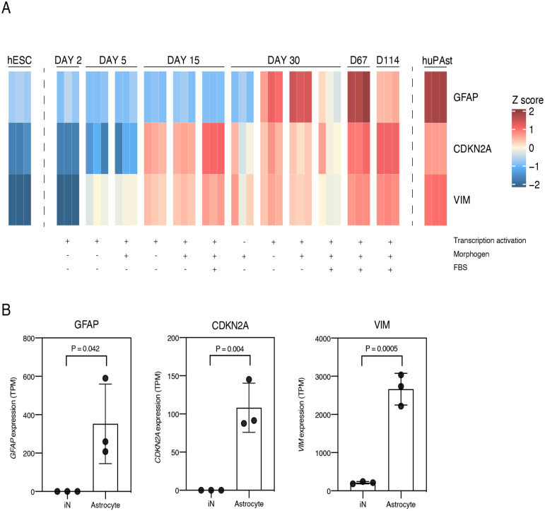
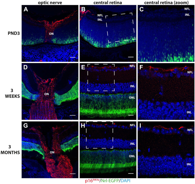
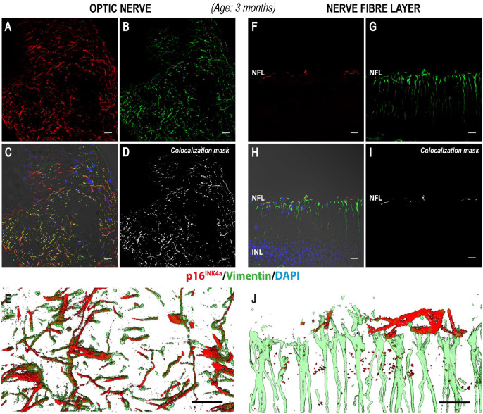
Retinal astrocytes are vital for neuronal homeostasis in the retina. Together with Müller glia, they provide retinal cells with neurotrophic factors, antioxidative support, and defense mechanisms such as the formation of the blood-retinal barrier. Substantial heterogeneity of astrocyte morphology and function represents a challenge for identification of distinct subtypes which may be potential targets for therapeutic purposes. Hence, identification of novel markers of astrocyte subpopulations is highly relevant to better understand the molecular mechanisms involved in retinal development, homeostasis, and pathology. In this study, we observed that the cell cycle regulator, p16INK4a, is expressed in immature astrocytes in the mouse retina. Immunohistochemical analysis showed p16INK4a expression in the optic nerve of wild-type mice from 3 days to 3 months of age and in the nerve fiber layer of the adult mouse retina. Colocalization of p16INK4a expression and glial fibrillary acidic protein (immature/mature astrocyte marker) tends to decrease with age. However, colocalization of p16INK4a expression and vimentin (immature astrocyte marker) remains high in the optic nerve from the early postnatal period to adulthood. The observations from this study provide a valuable tool for further investigations of ocular astrocytes in the developing retina as well as in degenerative retinopathies.
期刊介绍:
Journal of Histochemistry & Cytochemistry (JHC) has been a pre-eminent cell biology journal for over 50 years. Published monthly, JHC offers primary research articles, timely reviews, editorials, and perspectives on the structure and function of cells, tissues, and organs, as well as mechanisms of development, differentiation, and disease. JHC also publishes new developments in microscopy and imaging, especially where imaging techniques complement current genetic, molecular and biochemical investigations of cell and tissue function. JHC offers generous space for articles and recognizing the value of images that reveal molecular, cellular and tissue organization, offers free color to all authors.


 分享
分享

 求助内容:
求助内容: 应助结果提醒方式:
 扫码关注我们
