Gen Murakami, Kwang Ho Cho, Kei Kitamura, Jose Francisco Rodríguez-Vázquez, Tatsuo Sato
{"title":"重访头外侧直肌:一项使用人类胎儿的组织学研究。","authors":"Gen Murakami, Kwang Ho Cho, Kei Kitamura, Jose Francisco Rodríguez-Vázquez, Tatsuo Sato","doi":"10.1007/s00276-023-03237-1","DOIUrl":null,"url":null,"abstract":"<p><strong>Background: </strong>Rectus capitis lateralis muscle (RCLM) is located at the border between the ventral and dorsal muscle groups, but the nerve topographical anatomy around the muscle is obscure.</p><p><strong>Materials and methods: </strong>We observed the RCLM in histological sections of 12 midterm and 10 near-term fetal heads (9-18 and 26-40 weeks of gestational age).</p><p><strong>Results: </strong>At midterm, the RCLM wrapped around the inferiorly protruding inferolateral corner of the cartilaginous occipital bone. The muscle was adjacent to, or even continued to, the intertransversarius muscle between the atlas and axis. At near-term, the jugular process of the occipital bone, that is, the RCLM upper insertion, was either cartilaginous or bony, depending on age. The process formed a collar supporting the internal jugular vein from the inferior side. Moreover, the muscle is tightly attached to or inserted into the venous wall itself. The cartilaginous jugular process was adjacent to Reichert's cartilage, and the uppermost muscle fibers passed through a narrow space between these cartilages. The RCLM appeared to accelerate the jugular process elongation, resulting in complete union of the occipital and temporal bones. The ventral ramus of the first cervical nerve passed between the RCLM and rectus capitis anterior muscle to reach the longus capitis muscle. No nerve passed between the RCLM and the obliquus capitis superior muscle (a muscle at the suboccipital triangle).</p><p><strong>Conclusion: </strong>The dorsoventral position of the RCLM seemed to correspond to the scalenus posterior muscle in a laminar arrangement of the cervical axial musculature.</p>","PeriodicalId":49296,"journal":{"name":"Surgical and Radiologic Anatomy","volume":" ","pages":"1483-1491"},"PeriodicalIF":1.2000,"publicationDate":"2023-11-01","publicationTypes":"Journal Article","fieldsOfStudy":null,"isOpenAccess":false,"openAccessPdf":"","citationCount":"0","resultStr":"{\"title\":\"Rectus capitis lateralis muscle revisited: a histological study using human fetuses.\",\"authors\":\"Gen Murakami, Kwang Ho Cho, Kei Kitamura, Jose Francisco Rodríguez-Vázquez, Tatsuo Sato\",\"doi\":\"10.1007/s00276-023-03237-1\",\"DOIUrl\":null,\"url\":null,\"abstract\":\"<p><strong>Background: </strong>Rectus capitis lateralis muscle (RCLM) is located at the border between the ventral and dorsal muscle groups, but the nerve topographical anatomy around the muscle is obscure.</p><p><strong>Materials and methods: </strong>We observed the RCLM in histological sections of 12 midterm and 10 near-term fetal heads (9-18 and 26-40 weeks of gestational age).</p><p><strong>Results: </strong>At midterm, the RCLM wrapped around the inferiorly protruding inferolateral corner of the cartilaginous occipital bone. The muscle was adjacent to, or even continued to, the intertransversarius muscle between the atlas and axis. At near-term, the jugular process of the occipital bone, that is, the RCLM upper insertion, was either cartilaginous or bony, depending on age. The process formed a collar supporting the internal jugular vein from the inferior side. Moreover, the muscle is tightly attached to or inserted into the venous wall itself. The cartilaginous jugular process was adjacent to Reichert's cartilage, and the uppermost muscle fibers passed through a narrow space between these cartilages. The RCLM appeared to accelerate the jugular process elongation, resulting in complete union of the occipital and temporal bones. The ventral ramus of the first cervical nerve passed between the RCLM and rectus capitis anterior muscle to reach the longus capitis muscle. No nerve passed between the RCLM and the obliquus capitis superior muscle (a muscle at the suboccipital triangle).</p><p><strong>Conclusion: </strong>The dorsoventral position of the RCLM seemed to correspond to the scalenus posterior muscle in a laminar arrangement of the cervical axial musculature.</p>\",\"PeriodicalId\":49296,\"journal\":{\"name\":\"Surgical and Radiologic Anatomy\",\"volume\":\" \",\"pages\":\"1483-1491\"},\"PeriodicalIF\":1.2000,\"publicationDate\":\"2023-11-01\",\"publicationTypes\":\"Journal Article\",\"fieldsOfStudy\":null,\"isOpenAccess\":false,\"openAccessPdf\":\"\",\"citationCount\":\"0\",\"resultStr\":null,\"platform\":\"Semanticscholar\",\"paperid\":null,\"PeriodicalName\":\"Surgical and Radiologic Anatomy\",\"FirstCategoryId\":\"3\",\"ListUrlMain\":\"https://doi.org/10.1007/s00276-023-03237-1\",\"RegionNum\":4,\"RegionCategory\":\"医学\",\"ArticlePicture\":[],\"TitleCN\":null,\"AbstractTextCN\":null,\"PMCID\":null,\"EPubDate\":\"2023/9/2 0:00:00\",\"PubModel\":\"Epub\",\"JCR\":\"Q3\",\"JCRName\":\"ANATOMY & MORPHOLOGY\",\"Score\":null,\"Total\":0}","platform":"Semanticscholar","paperid":null,"PeriodicalName":"Surgical and Radiologic Anatomy","FirstCategoryId":"3","ListUrlMain":"https://doi.org/10.1007/s00276-023-03237-1","RegionNum":4,"RegionCategory":"医学","ArticlePicture":[],"TitleCN":null,"AbstractTextCN":null,"PMCID":null,"EPubDate":"2023/9/2 0:00:00","PubModel":"Epub","JCR":"Q3","JCRName":"ANATOMY & MORPHOLOGY","Score":null,"Total":0}
Rectus capitis lateralis muscle revisited: a histological study using human fetuses.
Background: Rectus capitis lateralis muscle (RCLM) is located at the border between the ventral and dorsal muscle groups, but the nerve topographical anatomy around the muscle is obscure.
Materials and methods: We observed the RCLM in histological sections of 12 midterm and 10 near-term fetal heads (9-18 and 26-40 weeks of gestational age).
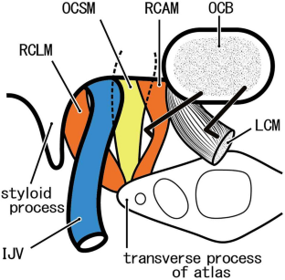
Results: At midterm, the RCLM wrapped around the inferiorly protruding inferolateral corner of the cartilaginous occipital bone. The muscle was adjacent to, or even continued to, the intertransversarius muscle between the atlas and axis. At near-term, the jugular process of the occipital bone, that is, the RCLM upper insertion, was either cartilaginous or bony, depending on age. The process formed a collar supporting the internal jugular vein from the inferior side. Moreover, the muscle is tightly attached to or inserted into the venous wall itself. The cartilaginous jugular process was adjacent to Reichert's cartilage, and the uppermost muscle fibers passed through a narrow space between these cartilages. The RCLM appeared to accelerate the jugular process elongation, resulting in complete union of the occipital and temporal bones. The ventral ramus of the first cervical nerve passed between the RCLM and rectus capitis anterior muscle to reach the longus capitis muscle. No nerve passed between the RCLM and the obliquus capitis superior muscle (a muscle at the suboccipital triangle).
Conclusion: The dorsoventral position of the RCLM seemed to correspond to the scalenus posterior muscle in a laminar arrangement of the cervical axial musculature.
期刊介绍:
Anatomy is a morphological science which cannot fail to interest the clinician. The practical application of anatomical research to clinical problems necessitates special adaptation and selectivity in choosing from numerous international works. Although there is a tendency to believe that meaningful advances in anatomy are unlikely, constant revision is necessary. Surgical and Radiologic Anatomy, the first international journal of Clinical anatomy has been created in this spirit.
Its goal is to serve clinicians, regardless of speciality-physicians, surgeons, radiologists or other specialists-as an indispensable aid with which they can improve their knowledge of anatomy. Each issue includes: Original papers, review articles, articles on the anatomical bases of medical, surgical and radiological techniques, articles of normal radiologic anatomy, brief reviews of anatomical publications of clinical interest.
Particular attention is given to high quality illustrations, which are indispensable for a better understanding of anatomical problems.
Surgical and Radiologic Anatomy is a journal written by anatomists for clinicians with a special interest in anatomy.


 分享
分享
 求助内容:
求助内容: 应助结果提醒方式:
 扫码关注我们
